. 2018 Sep 11;8(1):13607.
doi: 10.1038/s41598-018-31945-x.

The voltage-dependence of MscL has dipolar and dielectric contributions and is governed by local intramembrane electric field

Affiliations

The voltage-dependence of MscL has dipolar and dielectric contributions and is governed by local intramembrane electric field

Joseph S Najem et al. Sci Rep. .

Abstract

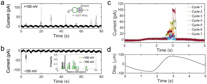
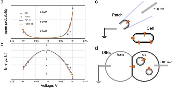
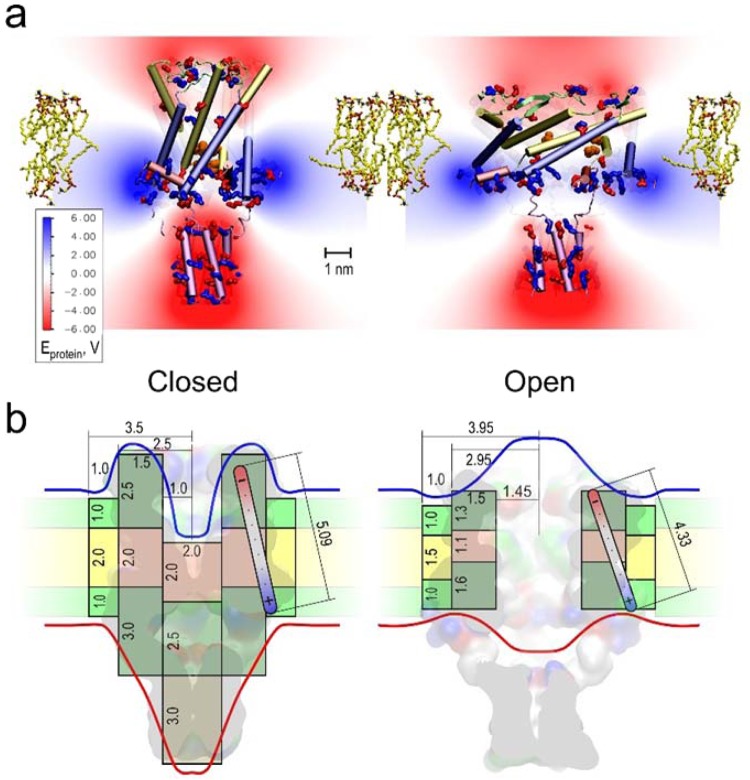
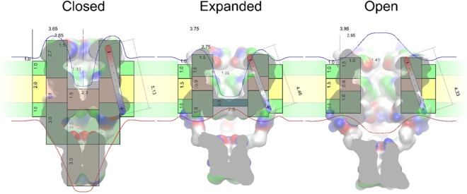
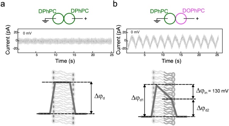
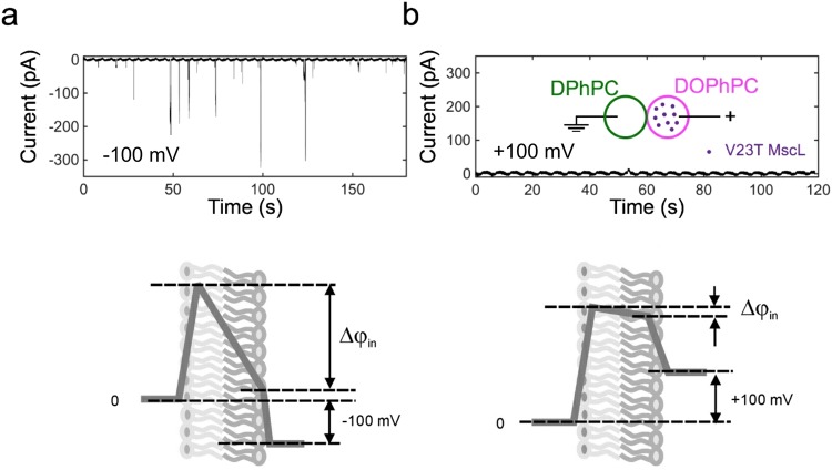
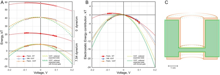
Channels without canonical voltage sensors can be modulated by voltage acting on other domains. Here we show that besides protein dipoles, pore hydration can be affected by electric fields. In patches, both WT MscL and its V23T mutant show a decrease in the tension midpoint with hyperpolarization. The mutant exhibits a stronger parabolic dependence of transition energy on voltage, highly consistent with the favourable dielectric contribution from water filling the expanding pore. Purified V23T MscL in DPhPC droplet interface bilayers shows a similar voltage dependence. When reconstituted in an asymmetric DOPhPC/DPhPC bilayer carrying a permanent bias of ~130 mV due to a dipole potential difference between the interfaces, the channel behaved as if the local intramembrane electric field sets the tension threshold for gating rather than just the externally applied voltage. The data emphasize the roles of polarized water in the pore and interfacial lipid dipoles in channel gating thermodynamics.


Conflict of interest statement

The authors declare no competing interests.

Figures

Figure 1
Figure 1
Voltage sensitivity of MscL. Current responses of WT MscL and V23T MscL populations to pressure ramps at different pipette voltages (imposed in 20 mV increments). Experiments were conducted in inside-out patches excised from giant bacterial spheroplasts expressing corresponding channels. Pressure midpoints for WT MscL (a) are higher at negative pipette voltages (asterisks), whereas V23T MscL (b) shows shallower and more symmetric responses and a more substantial shift to the left of the pressure midpoint with voltage. Both channels show ‘lingering’ conductive states at high negative voltages after stimulating pressure is released (black arrows). Plots of the closed-to-open transition energy extracted from activation curves as a function of pipette voltage (c,d). Parabolic fits predict different contributions from the capacitive (quadratic) and dipole (linear) components of the energy dependence on membrane potential, V (represented by right axes on panels c and d). This behaviour was reproduced in six independent patches for WT MscL and nine patches for V23T.
Figure 2
Figure 2
V23T MscL currents recorded in DIBs in the 0.75 duty cycle oscillating regime at different voltages. (a,b) Typical traces recorded at +100mV or −100 mV applied to the ‘cis’ compartment relative to the grounded ‘trans’ compartment. The distribution of observed conductances is shown in the inset in panel (b). (c) Seven segments of the continuous trace were recorded, each corresponding to one stimulation cycle (d) are overlaid to illustrate the position of opening events. As seen from panel (c), the channels open stochastically but tend to cluster around the 3 s time point, which is near the peak of compression where tension is highest in the membrane. In this particular trace, openings were observed in 21 out of 100 cycles.
Figure 3
Figure 3
Voltage dependences of open probability (a) and the closed-to-open transition energy (b) for V23T MscL recorded in DIBs (blue symbols) and in patch-clamp experiments (orange symbols). The patch data (orange) are simulated for the membrane tension at which the DIB data are collected. The fitting of open probability was done using Eq. 2. Based on the asymmetry of voltage response (a,b) we can deduce the orientation of the MscL channel incorporated in DIBs (c,d). In patches excised from the cytoplasmic membrane (c) the channel remains in its native orientation with its cytoplasmic side facing the bath. The directionality of MscL incorporation into DIBs is as presented in panel (d) and achieved through unilateral introduction of proteoliposomes into the ‘cis’ compartment.
Figure 4
Figure 4
Estimations of WT MscL changes in dipole and capacitive energies as a result of channel opening. Distributions of electric potential around the MscL complex in the closed (a) and open (b) conformations. The density of red and blue colour reflects the calculated electrostatic potential of the protein, negative to positive respectively. The opening transition changes the total dipole moment of the MscL complex from 2675 D (8.92∙10−27 C∙m) in the closed state to 2175 D (7.25∙10−27 C∙m) in the open state. Simplified cylindrical representations of the closed (c) and open (d) conformations of WT MscL superimposed with the molecular models of the closed and open states. Grey and pink regions represent high-dielectric (polar) and low-dielectric (apolar) segments of the protein; green and yellow represent polar and apolar regions of the annular lipids around the protein. Numbers represent thicknesses and radii of different dielectric segments that undergo conformational changes. The red and blue lines depict putative equipotential surfaces around the protein and membrane under positive voltage in the outer (periplasmic) compartment relative to the cytoplasm. The shape of equipotential surfaces reflects the redistribution of electric field around the closed and open pore.
Figure 5
Figure 5
Estimations of V23T MscL’s changes in dipole and capacitive energies as a result of transitions to the expanded (subconductive) and open states. Simplified cylindrical representations of three main conformations of V23T MscL (closed, expanded, and open) overlaid on atomistic models of respective states presented as surfaces. Grey regions represent high-dielectric (polar) and pink represent low-dielectric (apolar) segments of the protein; cyan domain in the expanded (subconductive) state shows a polar (likely hydrated) occlusion of the pore; green and yellow represent polar and apolar regions of the annular lipids around the protein. Numbers represent approximate thicknesses and diameters of different dielectric segments that undergo conformational changes in nm. The red and blue lines depict equipotential surfaces around the protein and membrane, in this particular case blue line designates positive voltage in the outer (periplasmic) compartment.
Figure 6
Figure 6
Displacement currents recorded on symmetric DPhPC/DPhPC and asymmetric DPhPC/DOPhPC DIBs in response to periodic mechanical oscillations. The external voltage was set to zero in both experiments, and the recordings were done in the absence of reconstituted channels.
Figure 7
Figure 7
Activities of V23T MscL reconstituted into an asymmetric DIB folded from DPhPC (left) and DOPhPC (right). Cartoons below depict electric field distribution across electrically neutral asymmetric membranes composed of two leaflets characterized by different surface dipole potentials. External voltage of −100 mV amplifies the internal potential Δφin, promoting massive gating of V23T MscL. Positive +100 mV potential negates the internal potential drop and V23T MscL remains silent.
Figure 8
Figure 8
The summary of voltage effects on the transition energy of WT (red), V23T (orange, blue) and a hypothetical mutant with a slightly wider barrel in the expanded state (light green) and also thinned gate (dark green) extrapolated to ±200 mV (pipette). The positive voltage in this plot corresponds to hyperpolarization. (A) Energies for the opening transition presented in the kT scale for WT and V23T at 0 tension (upper curves) and for the tension of 7.54 mN/m (lower curves). (B) The comparison of slopes of electrostatic contributions when the origins of all curves are rescaled to zero. (C) The cartoon illustrating the effective geometry of voltage-sensitive V23T MscL in the expanded subconductive state (orange) and the shape of a hypothetical re-engineered channel characterized by an open-like pore width and slightly thinned hydrophilic septum separating the compartments (green).

Similar articles

Cited by

References

    1. Bezanilla F. Voltage-gated ion channels. IEEE Trans Nanobioscience. 2005;4:34–48. doi: 10.1109/TNB.2004.842463. - DOI - PubMed
    1. Vargas E, et al. An emerging consensus on voltage-dependent gating from computational modeling and molecular dynamics simulations. J Gen Physiol. 2012;140:587–594. doi: 10.1085/jgp.201210873. - DOI - PMC - PubMed
    1. Anishkin A, Chiang CS, Sukharev S. Gain-of-function mutations reveal expanded intermediate states and a sequential action of two gates in MscL. The Journal of general physiology. 2005;125:155–170. doi: 10.1085/jgp.200409118. - DOI - PMC - PubMed
    1. Anishkin A, Akitake B, Kamaraju K, Chiang CS, Sukharev S. Hydration properties of mechanosensitive channel pores define the energetics of gating. J Phys Condens Matter. 2010;22:454120. doi: 10.1088/0953-8984/22/45/454120. - DOI - PubMed
    1. Sukharev SI, Sigurdson WJ, Kung C, Sachs F. Energetic and spatial parameters for gating of the bacterial large conductance mechanosensitive channel, MscL. The Journal of general physiology. 1999;113:525–540. doi: 10.1085/jgp.113.4.525. - DOI - PMC - PubMed

Publication types

LinkOut - more resources