. 2018 Dec 12;10(12):1728-1742.
doi: 10.1039/c8mt00133b.

Antimicrobial action of calprotectin that does not involve metal withholding

Affiliations

Antimicrobial action of calprotectin that does not involve metal withholding

Angelique N Besold et al. Metallomics. .

Abstract

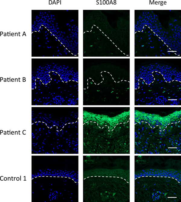
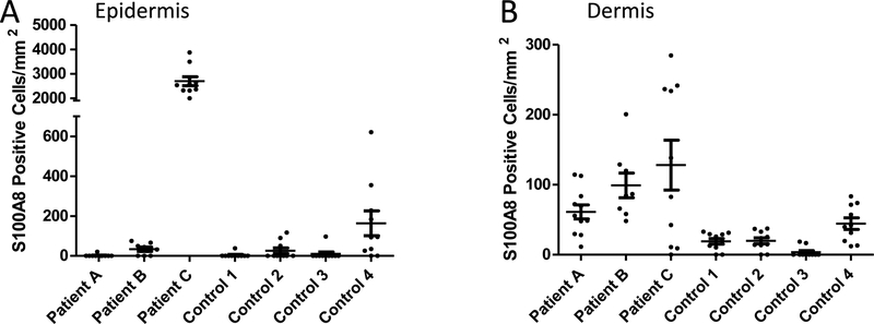
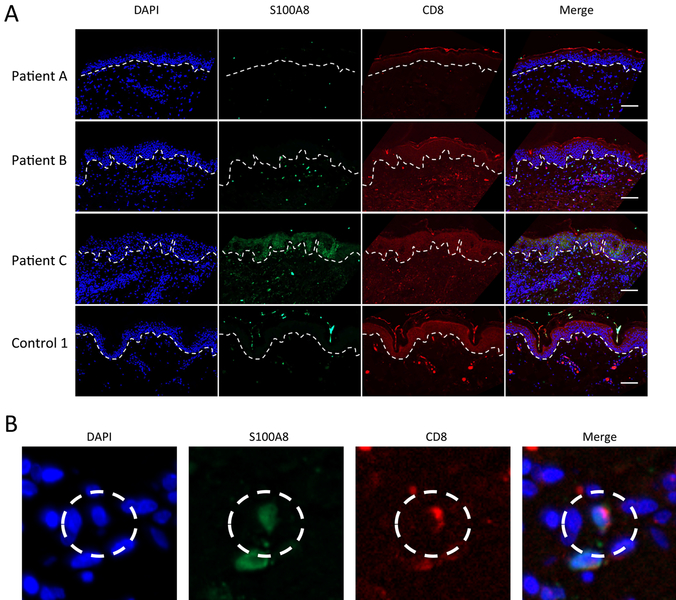
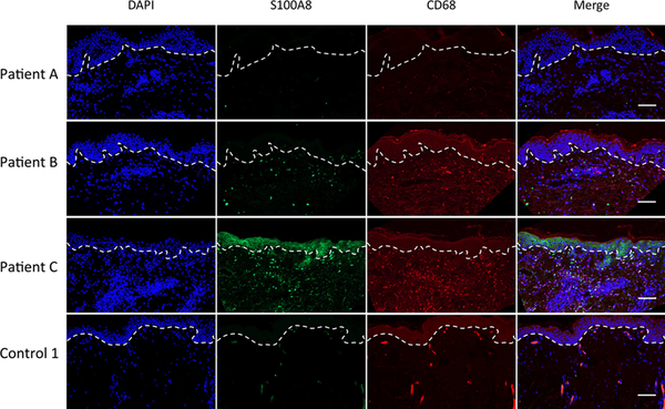
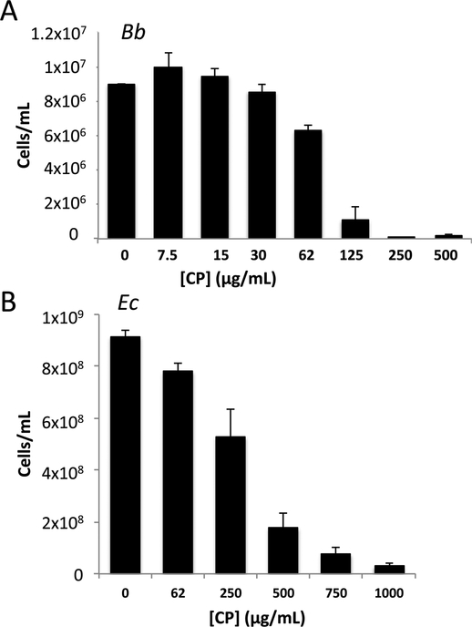
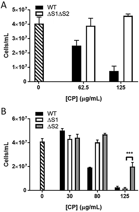
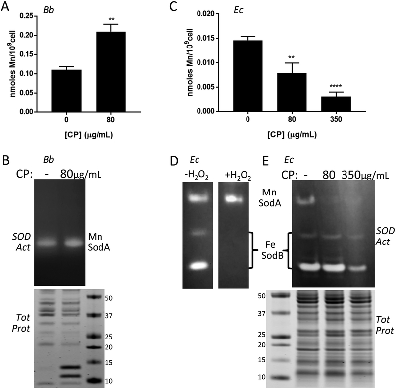
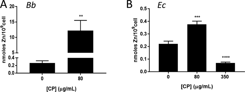
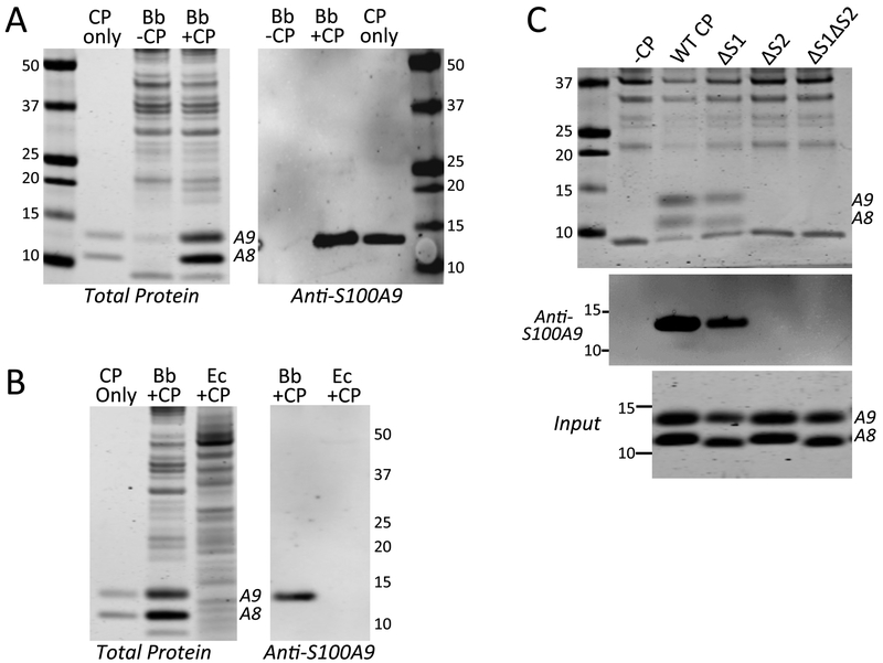
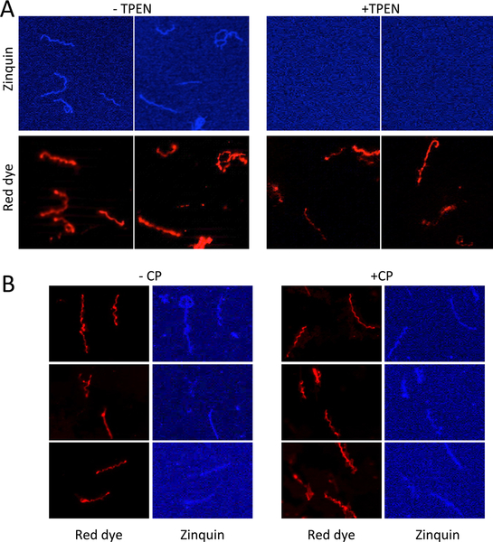
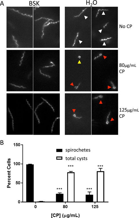
Calprotectin is a potent antimicrobial that inhibits the growth of pathogens by tightly binding transition metals such as Mn and Zn, thereby preventing their uptake and utilization by invading microbes. At sites of infection, calprotectin is abundantly released from neutrophils, but calprotectin is also present in non-neutrophil cell types that may be relevant to infections. We show here that in patients infected with the Lyme disease pathogen Borreliella (Borrelia) burgdorferi, calprotectin is produced in neutrophil-free regions of the skin, in both epidermal keratinocytes and in immune cells infiltrating the dermis, including CD68 positive macrophages. In culture, B. burgdorferi's growth is inhibited by calprotectin, but surprisingly, the mechanism does not involve the classical withholding of metal nutrients. B. burgdorferi cells exposed to calprotectin cease growth with no reduction in intracellular Mn and no loss in activity of Mn enzymes including the SodA superoxide dismutase. Additionally, there is no obvious loss in intracellular Zn. Rather than metal depletion, we find that calprotectin inhibits B. burgdorferi growth through a mechanism that requires physical association of calprotectin with the bacteria. By comparison, calprotectin inhibited E. coli growth without physically interacting with the microbe, and calprotectin effectively depleted E. coli of intracellular Mn and Zn. Our studies with B. burgdorferi demonstrate that the antimicrobial capacity of calprotectin is complex and extends well beyond simple withholding of metal micronutrients.


Conflict of interest statement

CONFLICTS OF INTEREST

There are no conflicts of interest to declare.

Figures

Fig. 1
Fig. 1
Immunofluorescence microscopy imaging of S100A8 expression in the skin of LD patients and controls. Skin tissue sections from the EM rash site of three individual LD patients and one control patient (Control 1) were subjected to immunostaining for S100A8 (green) and nuclei staining with DAPI (blue) before being subjected to fluorescence microscopy at 20× magnification as described in Experimental. Dotted lines separate the epidermis from the dermis. Bar represents 50 μm.
Fig. 2
Fig. 2
Quantification of S100A8 positive cells in the skin of LD patients. S100A8 positive cells were quantified in the epidermis (A) and dermis (B) of skin sections from 3 LD patients and 4 control surgical patients. Results represent cell counts (positive for both DAPI and S100A8) from 8–11 individual images spanning the tissue section of an individual patient. Image numbers: Patient A n=11; patient B n=8; patient C and all four controls n=10. (A) S100A8 staining in the epidermis of patient A is significantly higher than all four controls (p <0.001) and individual patients B and C (p<0.001). (B) S100A8 staining in the dermis of patient C is significantly higher than all four controls (p <0.01); the same is true for patient B and controls 1–3 (p<0.05).
Fig. 3
Fig. 3
Co-localization of S100A8 and CD8 expression in the dermis. (A) Skin tissue sections from the EM rash site of three individual LD patients and one control (control 1) were subjected to immunostaining for S100A8 (green), CD8 (red) and nuclei staining with DAPI (blue) before being subjected to fluorescence microscopy at 20× magnification as in Fig. 1. Dotted lines separate the epidermis from the dermis and bar represents 50 μm. (B) Individual cells co-expressing S100A8 and CD8 were identified by staining with anti-S100A8 (green), anti-CD68 (red) and DAPI (blue) as shown in the dotted circle.
Fig. 4
Fig. 4
Expression of S100A8 and CD68 expression in the dermis. Skin tissue section from the EM rash site of three individual LD patients and one control were subjected to immunostaining for S100A8 (green), CD68 (red) and nuclei staining with DAPI (blue) before being subjected to fluorescence microscopy at 20× magnification as in Fig. 1. Dotted lines separate the epidermis from the dermis and bar represents 50 μm.
Fig. 5
Fig. 5
Calprotectin (CP) mediated growth inhibition of B. burgdorferi (Bb) versus E. coli. Bb (A) or E. coli (B) were cultured in BSK II medium as described in Experimental in the presence of the indicated concentrations of CP. (A) Bb cell number was enumerated under dark field microscopy; results are averages of three biological replicates representative of seven experimental trials. There was essentially no growth at 250 and 500 μg/mL CP and the cell number approximated the original inoculum. (B) E. coli cell number was converted from optical density; results are the averages of four biological replicates and are representative of eight experimental trials. Error bars are standard error. Across numerous experimental trials, the mean inhibitory concentration or dose of CP that inhibits growth by 50% for Bb and E. coli were ≈80 and ≈350 μg/mL, respectively.
Fig. 6
Fig. 6
Effect of CP metal binding mutants on inhibiting growth of Bb. Bb cells were grown as in Fig. 5A in the presence of WT CP (black bar), the indicated metal binding mutants of CP (white and grey bars), or without CP (stippled bar). Results are averages of three biological replicates and are representative of five (A) and three (B) experimental trials; error bars are standard error. The difference in growth inhibition between ΔS1 and ΔS2 CP at 125 μg/mL is statistically significant; ***p = 0.001.
Fig. 7
Fig. 7
Effects of CP on Mn requiring SodA and total cellular Mn in Bb versus E. coli. Bb (A, B) and E. coli cells (“Ec” C-E) were grown in BSK II supplemented with the designated concentrations of CP. (A,C) Mn levels were measured in Bb (A) or E. coli (C) cells by ICP-MS. Results are averages of at least five (A) and four (C) replicates over three experimental trials, **p ≤ 0.0035, ****p < 0.0001, where CP treated samples are compared to no CP controls. Error bars are standard error. (B,D,E) Whole cell lysates were analyzed for SOD enzymatic activity by native gel electrophoresis and NBT staining (B top, D, E top). Denaturing gel electrophoresis and coomassie staining (B bottom, E bottom) were used as a loading control. The positions of SodA and SodB on the native gels are indicated; numbers represent molecular weight markers. Results are representative of five (B) and three (E) experimental trials. (D) The native gel was treated with H2O2 where indicated to inactivate Fe containing SodB prior to NBT staining, as described in Experimental.
Fig. 8
Fig. 8
Zn and CP in Bb versus E. coli. Total Zn levels were measured by ICP-MS in whole cell samples of Bb or E. coli grown with the indicated concentration of CP. (A) Results are averages of eight (0 μg/mL CP) and eleven (80 μg/mL CP) replicates over five independent experiments, where the level of Zn in CP treated cell samples ranged from ≈6–30 nmoles/109 cells; **p=0.0073. (B) Results are averages of 10 (0 μg/mL CP), 8 (80 μg/mL CP), and 7 (350 μg/mL CP) replicates over four experimental trials, ***p=0.0004; ****p<0.0001.
Fig. 9
Fig. 9
Interactions between CP and Bb. Bb or E. coli cells were grown in BSK II in the absence or presence of 80 or 350 μg/mL CP, (A, B). Bb was grown with 80 μg/mL of the designated CP mutants (C). Whole cell lysates were subjected to denaturing gel electrophoresis and either coomassie staining for total protein (A,B left, C top) or immunoblot analysis for S100A9 (“Anti-S100A9”). “CP only” is 500 ng of recombinant human CP. Numbers indicate molecular weight markers and positions of S100A8 and S100A9 are denoted. “Input” shows coomassie staining of WT and the indicated CP mutants added to Bb cultures. Over five experimental trials, the level of ΔS1 CP recovered in Bb lysates (as in Fig. 9C top and middle panels) was 42% (± 12%) that of WT CP, while ΔS2 and ΔS1ΔS2 CP were undetected.
Fig. 10
Fig. 10
Zinquin labeling of Bb. Bb cells were cultured in the absence (A, left) or presence (A, right) of 3 μM TPEN, a Zn chelator, or in the absence (B, left) or presence (B, right) of 80 μg/mL CP. Cells were prepared for fluorescence microscopy by sequential staining with zinquin and then PKH red dye (for membranes) as described in Experimental. Results are representative of 10 (A) and 20 (B) images.
Fig. 11:
Fig. 11:
CP and morphology of Bb. (A) Bb cells cultured with the indicated concentrations of CP were examined by dark field microscopy either directly in BSK II medium or immediately following a 1:10 dilution in H2O. Arrows indicate morphology classes: white, elongated spirals; red, cysts with round body tip; yellow, full round body cysts. Similar results were obtained with 1:5 dilution in H2O (Fig. S5, ESI†). (B) Quantification of cell morphology following dilution in H2O. Cysts are a combination of the two classes described above where full round bodies are less <5% total cysts. Results represent the averages of >250 cells counted over 3–4 experimental trials. Error bars are standard error. The changes in morphology with 80 and 125 μg/mL CP compared to no CP are statistically significant; ***p<0.001

References

    1. Hood MI and Skaar EP, Nutritional immunity: transition metals at the pathogen-host interface, Nat. Rev. Microbiol, 2012, 10, 525–537. - PMC - PubMed
    1. Zackular JP, Chazin WJ and Skaar EP, Nutritional Immunity: S100 Proteins at the Host-Pathogen Interface, J. Biol. Chem, 2015, 290, 18991–18998. - PMC - PubMed
    1. Zygiel EM and Nolan EM, Transition Metal Sequestration by the Host-Defense Protein Calprotectin, Annu. Rev. Biochem, 2018, 87, 621–643. - PMC - PubMed
    1. Vogl T, Leukert N, Barczyk K, Strupat K and Roth J, Biophysical characterization of S100A8 and S100A9 in the absence and presence of bivalent cations, Biochim. Biophys. Acta, 2006, 1763, 1298–1306. - PubMed
    1. Strupat K, Rogniaux H, Van Dorsselaer A, Roth J and Vogl T, Calcium-induced noncovalently linked tetramers of MRP8 and MRP14 are confirmed by electrospray ionization-mass analysis, J. Am. Soc. Mass Spectrom, 2000, 11, 780–788. - PubMed

Publication types

MeSH terms