

Glomangiopericytoma of the Nasal Cavity with CTNNB1 p.S37C Mutation: A Case Report and Literature Review
- PMID: 30206803
- PMCID: PMC6684555
- DOI: 10.1007/s12105-018-0961-z
Glomangiopericytoma of the Nasal Cavity with CTNNB1 p.S37C Mutation: A Case Report and Literature Review
Abstract
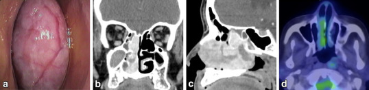
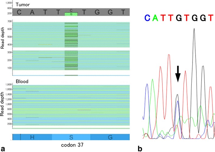
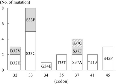
Glomangiopericytoma (GPC) is a rare mesenchymal tumor arising from the nasal cavity or paranasal sinuses. GPC was categorized as a borderline and low-malignant-potential tumor by the World Health Organization in 2005 and accounts for less than 0.5% of all sinonasal tumors. We report a case of GPC in a 74-year-old woman with a history of recurrent epistaxis and nasal obstruction. A reddish tumor was seen in the right nasal cavity. Enhanced computed tomography showed a mass lesion occupying the right nasal cavity. The tumor, which originated from the nasal septum in the olfactory fissure area, was resected with 5-mm mucosal margins by endoscopic sinus surgery. Histologic examination revealed a uniform proliferation of oval-to-short spindle-shaped cells beneath the epithelium. Immunohistologic analysis demonstrated the tumor cells were positive for α-smooth muscle actin, β-catenin and Vimentin, and negative for AE1/AE3, Bcl-2, CD34, CD117, Factor VIIIR Ag, S-100 protein, or STAT6. The percentage of Ki-67-positive cells was approximately 5%. Genetic analysis using next-generation sequencing revealed a missense mutation in the CTNNB1 gene (c.110C > G, p.S37C). While other CTNNB1 mutations have been described in GPC; this is the first report of this specific mutation. The mutation was confirmed using Sanger sequencing.
Keywords: CTNNB1; Endoscopic sinus surgery (ESS); Glomangiopericytoma (GPC); Next-generation sequencing (NGS).
Conflict of interest statement
The authors declare that there are no actual or potential conflicts of interest in relation to this article.
Figures

References
-
- Thompson LD, Fanburg-Smith J, Wenig B. Tumours of the nasal cavity and paranasal sinuses. Borderline and low malignant potential tumours of soft tissue. In: Barnes L, Eveson JW, Reichart P, Sidransky D, editors. World Health Organization (WHO) classification of tumours. Vol.9. Pathology and genetics of head and neck tumours. Lyon: IARC Press; 2005. pp. 43–44.
-
- Dandekar M, McHugh JB. Sinonasal glomangiopericytoma: case report with emphasis on the differential diagnosis. Arch Pathol Lab Med. 2010;134:1444–1449. - PubMed
Publication types
MeSH terms
Substances
LinkOut - more resources
Full Text Sources
Other Literature Sources
Medical
Research Materials
Miscellaneous
