. 2017 Nov 2;33(2):198-212.
doi: 10.5606/ArchRheumatol.2018.6519. eCollection 2018 Jun.

Bee Venom and Hesperidin Effectively Mitigate Complete Freund's Adjuvant-Induced Arthritis Via Immunomodulation and Enhancement of Antioxidant Defense System

Affiliations

Bee Venom and Hesperidin Effectively Mitigate Complete Freund's Adjuvant-Induced Arthritis Via Immunomodulation and Enhancement of Antioxidant Defense System

Osama Ahmed et al. Arch Rheumatol. .

Abstract

Objectives: This study aims to assess the antirheumatic activity of bee venom (BV) and/or hesperidin as natural products in complete Freund's adjuvant (CFA)-induced arthritis in male Wistar rats.

Material and methods: Rheumatoid arthritis was induced in 30 male Wistar rats (weight 130 g to 150 g; age 10 to 12 weeks) by subcutaneous injection of CFA into the right hind paw of the rats. The rats were divided into five groups of six rats in each and administered the following regimens for 21 days: Normal group (given the equivalent volume of saline and carboxymethylcellulose), arthritic group (given the equivalent volume of saline and carboxymethylcellulose), arthritic group treated with BV (treated with BV along with carboxymethylcellulose), arthritic group treated with hesperidin (treated with hesperidin along with saline), and arthritic group treated with BV and hesperidin (treated with BV and hesperidin concurrently).

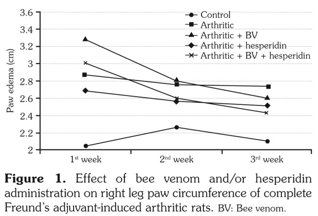
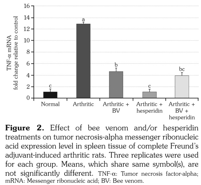
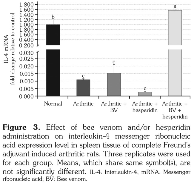
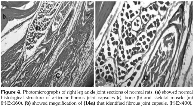
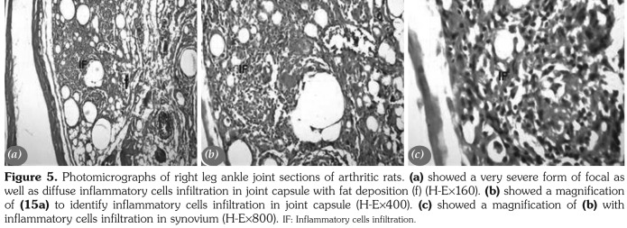
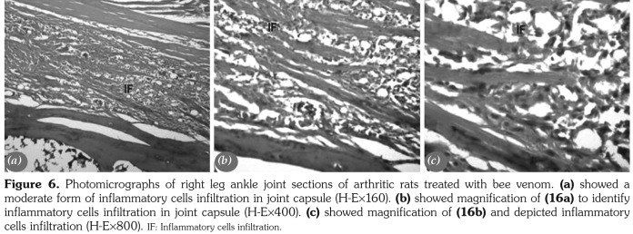
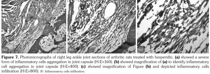
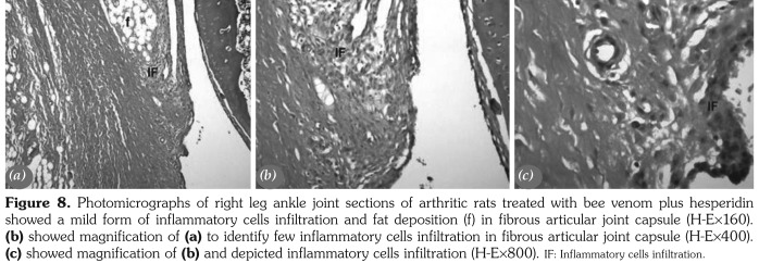
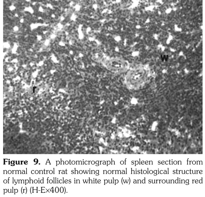
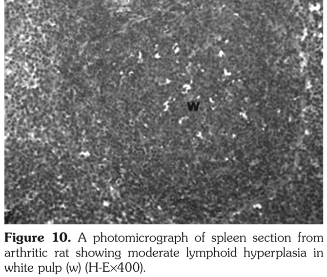
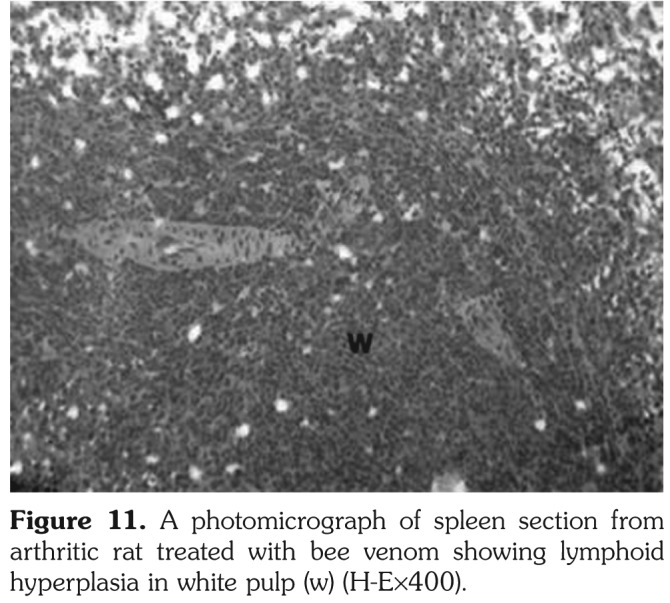
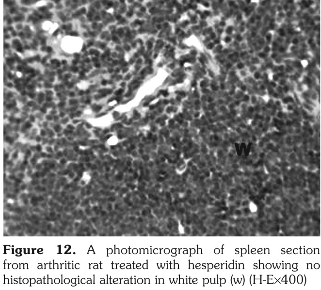
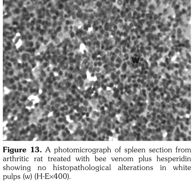
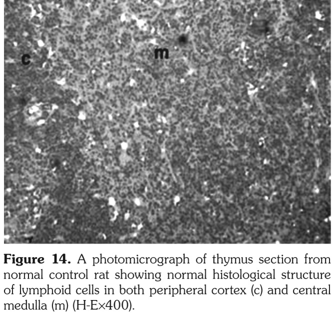
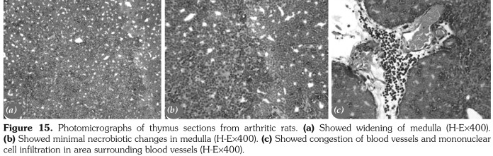
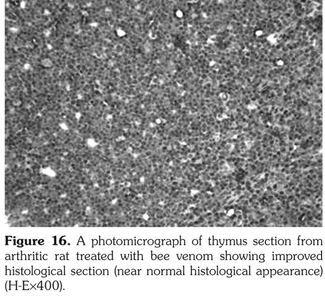
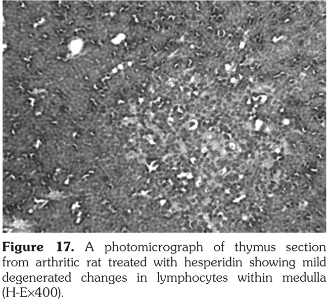
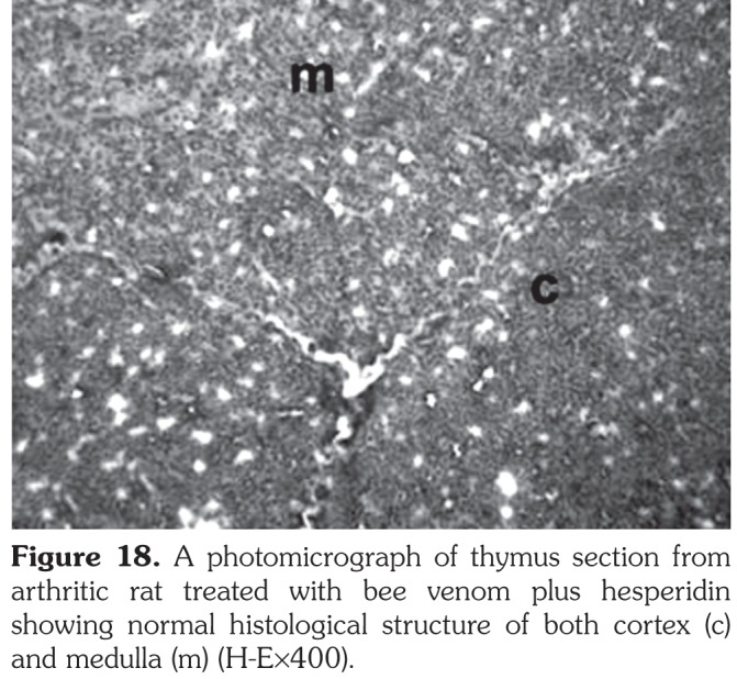
Results: Bee venom and/or hesperidin successfully reversed the CFA-arthritis-induced increases in right hind leg paw swelling, leukocytes' count, liver lipid peroxidation, serum inflammatory cytokine interleukin (IL-2 and IL-12) levels and spleen tumor necrosis factor-alpha messenger ribonucleic acid expression. Moreover, the CFA-induced down-regulation in serum IL-10 level and spleen IL-4 messenger ribonucleic acid expression as well as the deterioration in the antioxidant defense system were significantly improved as a result of BV and hesperidin administration. Both treatments also markedly counteracted the severe inflammatory changes and leukocytic infiltration in the periarticular tissue of the ankle joints. In addition, BV and hesperidin obviously amended the lymphoid hyperplasia in white pulps of spleen as well as the widening of the medulla and mononuclear cell infiltration found in thymus.

Conclusion: Bee venom and hesperidin administration produced their ameliorative effects on rheumatoid arthritis via their antioxidant, antiinflammatory and immunomodulatory potentials. BV plus hesperidin particularly seemed to be the most potent in improving rheumatoid arthritis in Wistar rats.

Keywords: Bee venom; hesperidin; inflammation; oxidative stress; rheumatoid arthritis.


Conflict of interest statement

Conflict of Interest: The authors declared no conflicts of interest with respect to the authorship and/or publication of this article.

Figures

Figure 1
Figure 1. Effect of bee venom and/or hesperidin administration on right leg paw circumference of complete Freund’s adjuvant-induced arthritic rats. BV: Bee venom.
Figure 2
Figure 2. Effect of bee venom and/or hesperidin treatments on tumor necrosis-alpha messenger ribonucleic acid expression level in spleen tissue of complete Freund’s adjuvant-induced arthritic rats. Three replicates were used for each group. Means, which share same symbol(s), are not significantly different. TNF-a: Tumor necrosis factor-alpha; mRNA: Messenger ribonucleic acid; BV: Bee venom.
Figure 3
Figure 3. Effect of bee venom and/or hesperidin administration on interleukin-4 messenger ribonucleic acid expression level in spleen tissue of complete Freund's adjuvant-induced arthritic rats. Three replicates were used for each group. Means, which share same symbol(s), are not significantly different. IL-4: Interleukin-4; mRNA: Messenger ribonucleic acid; BV: Bee venom.
Figure 4
Figure 4. Photomicrographs of right leg ankle joint sections of normal rats. (a) showed normal histological structure of articular fibrous joint capsules (c), bone (b) and skeletal muscle (m) (H-Ex160). (b) showed magnification of (14a) that identified fibrous joint capsule. (H-Ex400).
Figure 5
Figure 5. Photomicrographs of right leg ankle joint sections of arthritic rats. (a) showed a very severe form of focal as well as diffuse inflammatory cells infiltration in joint capsule with fat deposition (f) (H-Ex160). (b) showed a magnification of (15a) to identify inflammatory cells infiltration in joint capsule (H-Ex400). (c) showed a magnification of (b) with inflammatory cells infiltration in synovium (H-Ex800). IF: Inflammatory cells infiltration.
Figure 6
Figure 6. Photomicrographs of right leg ankle joint sections of arthritic rats treated with bee venom. (a) showed a moderate form of inflammatory cells infiltration in joint capsule (H-Ex160). (b) showed magnification of (16a) to identify inflammatory cells infiltration in joint capsule (H-Ex400). (c) showed magnification of (16b) and depicted inflammatory cells infiltration (H-Ex800). IF: Inflammatory cells infiltration.
Figure 7
Figure 7. Photomicrographs of right leg ankle joint sections of arthritic rats treated with hesperidin. (a) showed a severe form of inflammatory cells aggregation in joint capsule (H-Ex160). (b) showed magnification of (a) to identify inflammatory cell aggregation in joint capsule (H-Ex400). (c) showed magnification of Figure (b) and depicted inflammatory cells infiltration (H-Ex800). IF: Inflammatory cells infiltration.
Figure 8
Figure 8. Photomicrographs of right leg ankle joint sections of arthritic rats treated with bee venom plus hesperidin showed a mild form of inflammatory cells infiltration and fat deposition (f) in fibrous articular joint capsule (H-Ex160). (b) showed magnification of (a) to identify few inflammatory cells infiltration in fibrous articular joint capsule (H-Ex400). (c) showed magnification of (b) and depicted inflammatory cells infiltration (H-Ex800). IF: Inflammatory cells infiltration.
Figure 9
Figure 9. A photomicrograph of spleen section from normal control rat showing normal histological structure of lymphoid follicles in white pulp (w) and surrounding red pulp (r) (H-Ex400).
Figure 10
Figure 10. A photomicrograph of spleen section from arthritic rat showing moderate lymphoid hyperplasia in white pulp (w) (H-Ex400).
Figure 11
Figure 11. A photomicrograph of spleen section from arthritic rat treated with bee venom showing lymphoid hyperplasia in white pulp (w) (H-Ex400).
Figure 12
Figure 12. A photomicrograph of spleen section from arthritic rat treated with hesperidin showing no histopathological alteration in white pulp (w) (H-Ex400)
Figure 13
Figure 13. A photomicrograph of spleen section from arthritic rat treated with bee venom plus hesperidin showing no histopathological alterations in white pulps (w) (H-Ex400).
Figure 14
Figure 14. A photomicrograph of thymus section from normal control rat showing normal histological structure of lymphoid cells in both peripheral cortex (c) and central medulla (m) (H-Ex400).
Figure 15
Figure 15. Photomicrographs of thymus sections from arthritic rats. (a) Showed widening of medulla (H-Ex400). (b) Showed minimal necrobiotic changes in medulla (H-Ex400). (c) Showed congestion of blood vessels and mononuclear cell infiltration in area surrounding blood vessels (H-Ex400).
Figure 16
Figure 16. A photomicrograph of thymus section from arthritic rat treated with bee venom showing improved histological section (near normal histological appearance) (H-Ex400).
Figure 17
Figure 17. A photomicrograph of thymus section from arthritic rat treated with hesperidin showing mild degenerated changes in lymphocytes within medulla (H-Ex400).
Figure 18
Figure 18. A photomicrograph of thymus section from arthritic rat treated with bee venom plus hesperidin showing normal histological structure of both cortex (c) and medulla (m) (H-Ex400).

References

    1. Imboden JB. The immunopathogenesis of rheumatoid arthritis. Annu Rev Pathol. 2009;4:417–434. - PubMed
    1. Brooks PM. The burden of musculoskeletal disease--a global perspective. Clin Rheumatol. 2006;25:778–781. - PubMed
    1. Zhang ZC, Zhang SJ, Jin B, Wu Y, Yang XF, Yu B, et al. Ciclamilast ameliorates adjuvant- induced arthritis in a rat model. Biomed Res Int. 2015;2015:786104–786104. - PMC - PubMed
    1. Firestein GS. Kelley’s textbook of rheumatology. 9. Etiology and pathogenesis of rheumatoid arthritis; p. p.
    1. Scott DL, Wolfe F, Huizinga TW. Rheumatoid arthritis. Lancet. 2010;376:1094–1108. - PubMed

LinkOut - more resources