

Cortico-cerebellar network involved in saccade adaptation
- PMID: 30207858
- PMCID: PMC6295533
- DOI: 10.1152/jn.00392.2018
Cortico-cerebellar network involved in saccade adaptation
Abstract
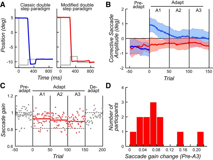
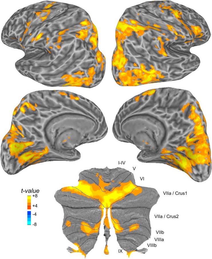
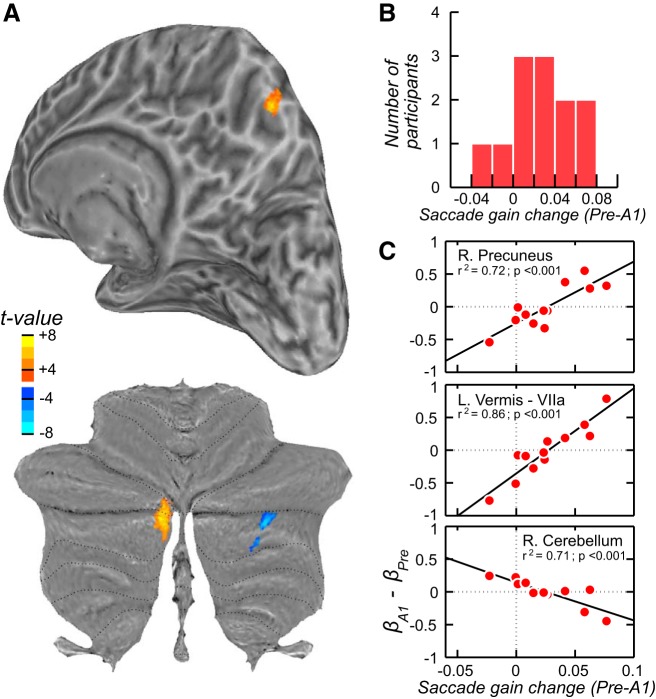
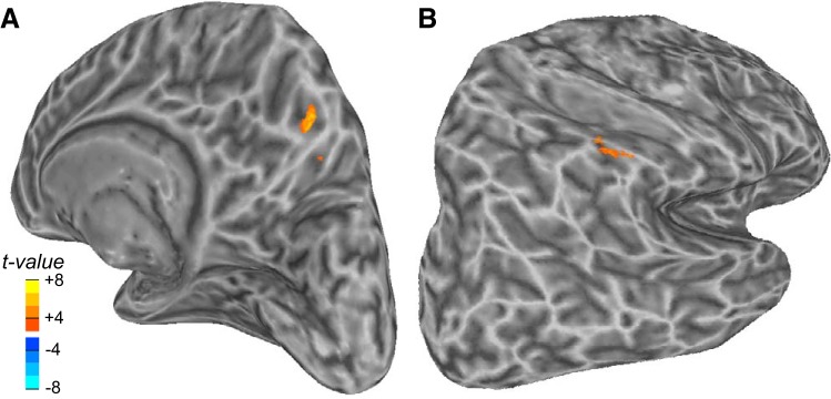
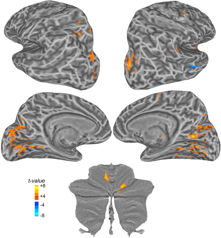
Saccade adaptation is the learning process that ensures that vision and saccades remain calibrated. The central nervous system network involved in these adaptive processes remains unclear because of difficulties in isolating the learning process from the correlated visual and motor processes. Here we imaged the human brain during a novel saccade adaptation paradigm that allowed us to isolate neural signals involved in learning independent of the changes in the amplitude of corrective saccades usually correlated with adaptation. We show that the changes in activation in the ipsiversive cerebellar vermis that track adaptation are not driven by the changes in corrective saccades and thus provide critical supporting evidence for previous findings. Similarly, we find that activation in the dorsomedial wall of the contraversive precuneus mirrors the pattern found in the cerebellum. Finally, we identify dorsolateral and dorsomedial cortical areas in the frontal and parietal lobes that encode the retinal errors following inaccurate saccades used to drive recalibration. Together, these data identify a distributed network of cerebellar and cortical areas and their specific roles in oculomotor learning. NEW & NOTEWORTHY The central nervous system constantly learns from errors and adapts to keep visual targets and saccades in registration. We imaged the human brain while the gain of saccades adapted to a visual target that was displaced while the eye was in motion, inducing retinal error. Activity in the cerebellum and precuneus tracked learning, whereas parts of the dorsolateral and dorsomedial frontal and parietal cortex encoded the retinal error used to drive learning.
Keywords: cerebellum; motor learning; oculomotor vermis; precuneus; saccade adaptation.
Figures
Similar articles
-
Changes in simple spike activity of some Purkinje cells in the oculomotor vermis during saccade adaptation are appropriate to participate in motor learning.J Neurosci. 2010 Mar 10;30(10):3715-27. doi: 10.1523/JNEUROSCI.4953-09.2010. J Neurosci. 2010. PMID: 20220005 Free PMC article.
-
Complex spike activity in the oculomotor vermis of the cerebellum: a vectorial error signal for saccade motor learning?J Neurophysiol. 2008 Oct;100(4):1949-66. doi: 10.1152/jn.90526.2008. Epub 2008 Jul 23. J Neurophysiol. 2008. PMID: 18650308 Free PMC article.
-
Effects of lesions of the oculomotor vermis on eye movements in primate: saccades.J Neurophysiol. 1998 Oct;80(4):1911-31. doi: 10.1152/jn.1998.80.4.1911. J Neurophysiol. 1998. PMID: 9772249
-
How cerebellar motor learning keeps saccades accurate.J Neurophysiol. 2019 Jun 1;121(6):2153-2162. doi: 10.1152/jn.00781.2018. Epub 2019 Apr 17. J Neurophysiol. 2019. PMID: 30995136 Free PMC article. Review.
-
Cortical control of saccades.Exp Brain Res. 1998 Nov;123(1-2):159-63. doi: 10.1007/s002210050557. Exp Brain Res. 1998. PMID: 9835405 Review.
Cited by
-
Saccadic adaptation shapes perceived size: Common codes for action and perception.Atten Percept Psychophys. 2020 Oct;82(7):3676-3685. doi: 10.3758/s13414-020-02102-2. Atten Percept Psychophys. 2020. PMID: 32725486
-
Adaptation across the 2D population code explains the spatially distributive nature of motor learning.PLoS Comput Biol. 2025 Jun 4;21(6):e1013041. doi: 10.1371/journal.pcbi.1013041. eCollection 2025 Jun. PLoS Comput Biol. 2025. PMID: 40465590 Free PMC article.
-
Visuomotor learning from postdictive motor error.Elife. 2021 Mar 9;10:e64278. doi: 10.7554/eLife.64278. Elife. 2021. PMID: 33687328 Free PMC article.
-
A triple distinction of cerebellar function for oculomotor learning and fatigue compensation.PLoS Comput Biol. 2023 Aug 4;19(8):e1011322. doi: 10.1371/journal.pcbi.1011322. eCollection 2023 Aug. PLoS Comput Biol. 2023. PMID: 37540726 Free PMC article.
-
Reactive saccade adaptation boosts orienting of visuospatial attention.Sci Rep. 2020 Aug 10;10(1):13430. doi: 10.1038/s41598-020-70120-z. Sci Rep. 2020. PMID: 32778710 Free PMC article.
References
Publication types
MeSH terms
Grants and funding
LinkOut - more resources
Full Text Sources
Other Literature Sources