. 2018 Dec 1;120(6):3026-3041.
doi: 10.1152/jn.00483.2018. Epub 2018 Sep 12.

Vestibular and corticospinal control of human body orientation in the gravitational field

Affiliations

Vestibular and corticospinal control of human body orientation in the gravitational field

Lei Zhang et al. J Neurophysiol. .

Abstract

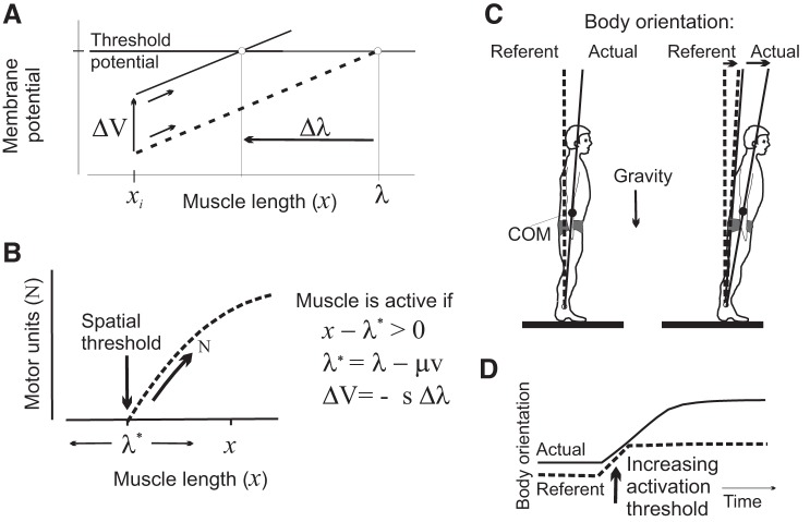
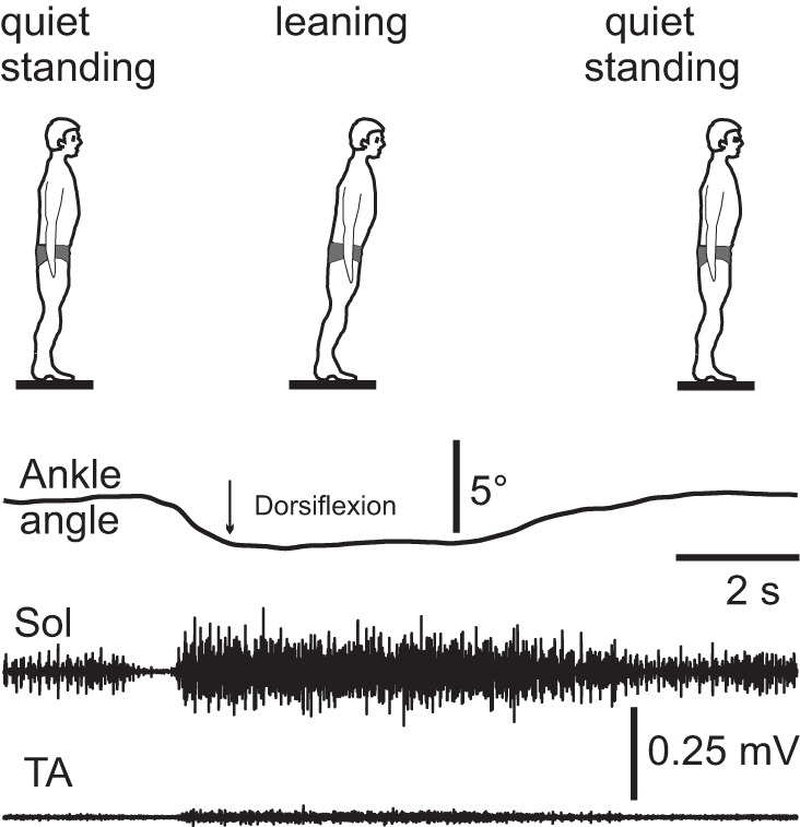
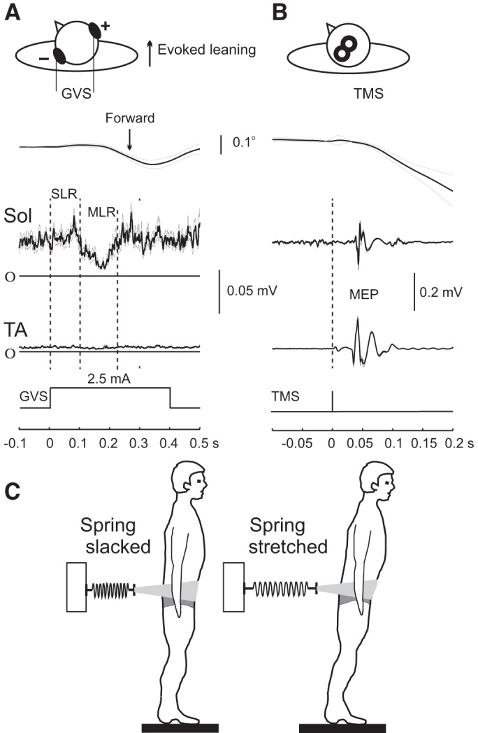
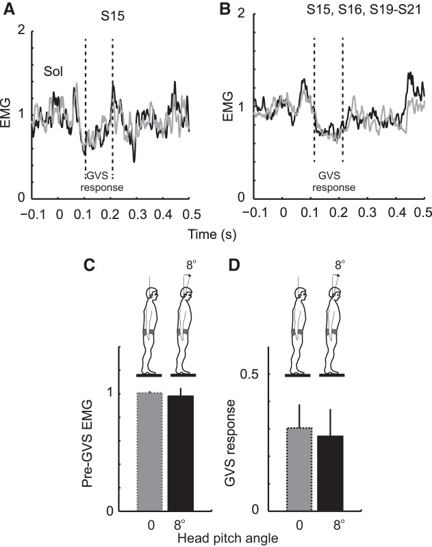
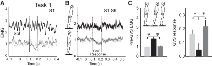
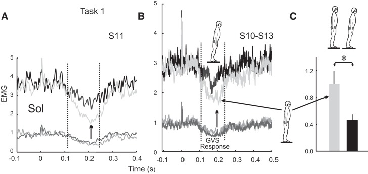
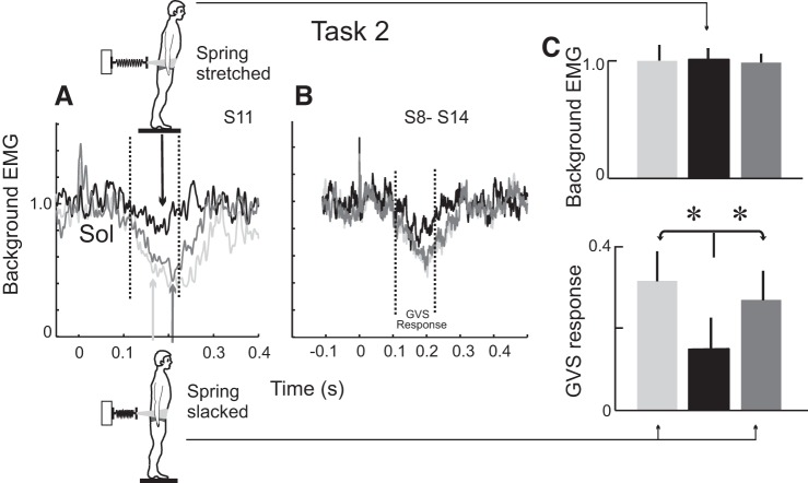
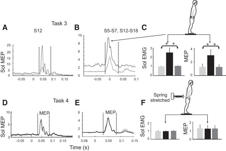
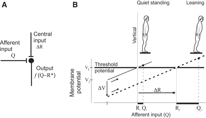
Body orientation with respect to the direction of gravity changes when we lean forward from upright standing. We tested the hypothesis that during upright standing, the nervous system specifies the referent body orientation that defines spatial thresholds for activation of multiple muscles across the body. To intentionally lean the body forward, the system is postulated to transfer balance and stability to the leaned position by monotonically tilting the referent orientation, thus increasing the activation thresholds of ankle extensors and decreasing their activity. Consequently, the unbalanced gravitational torque would start to lean the body forward. With restretching, ankle extensors would be reactivated and generate increasing electromyographic (EMG) activity until the enhanced gravitational torque would be balanced at a new posture. As predicted, vestibular influences on motoneurons of ankle extensors evaluated by galvanic vestibular stimulation were smaller in the leaned compared with the upright position, despite higher tonic EMG activity. Defacilitation of vestibular influences was also observed during forward leaning when the EMG levels in the upright and leaned position were equalized by compensating the gravitational torque with a load. The vestibular system is involved in the active control of body orientation without directly specifying the motor outcome. Corticospinal influences originating from the primary motor cortex evaluated by transcranial magnetic stimulation remained similar at the two body postures. Thus, in contrast to the vestibular system, the corticospinal system maintains a similar descending facilitation of motoneurons of leg muscles at different body orientations. The study advances the understanding of how body orientation is controlled. NEW & NOTEWORTHY The brain changes the referent body orientation with respect to gravity to lean the body forward. Physiologically, this is achieved by shifts in spatial thresholds for activation of ankle muscles, which involves the vestibular system. Results advance the understanding of how the brain controls body orientation in the gravitational field. The study also extends previous evidence of empirical control of motor function, i.e., without the reliance on model-based computations and direct specification of motor outcome.

Keywords: GVS; TMS; corticospinal; leaning; movement; posture; referent control; vestibular.


Figures

Fig. 1.
Fig. 1.
Referent control of motoneurons and body orientation in the gravitational field. A: because of length-dependent afferent feedback, the membrane potential of the α-motoneuron (α-MN) increases (dashed diagonal line) when the muscle is stretched from an initial length (xi). At a specific length called the threshold muscle length (λ), the potential exceeds the electrical threshold and the MN begins to be recruited. Central, length-independent changes in the membrane potential of α-MNs (ΔV; vertical arrow) elicited by direct and/or indirect influences (via interneurons and γ-MNs) result in a shift (Δλ) in the spatial activation threshold (horizontal arrow): ΔV = −sΔλ (s is sensitivity of the α-MN to changes in the muscle length). Thus the primary measure of changes in influences (facilitation and/or inhibition) on α-MNs is a shift in the spatial activation threshold. When the muscle is stretched at velocity v, activation threshold λ* decreases. Sensitivity (μ) of λ* to v can be regulated by γ-dynamic MNs. B: motor units of a given muscle begin to be recruited when the difference between the actual (x) and the dynamic threshold muscle length (λ*) becomes positive. The number of recruited motor units (N) and the muscle force increase (diagonal arrow) with this difference (x − λ*). C: actual orientation of the body relative to the vertical is the slope of the vector along the body from the ankle joint (solid line). Referent orientation (dashed line) is the body orientation at which multiple muscles reach their activation thresholds (or produce net zero torque if muscles are coactivated). It is assumed that during upright standing (left), the referent orientation is aligned with the vertical. Under the influence of gravitational torques, the body leans somewhat forward to an actual orientation at which body balance is achieved. To intentionally lean the body forward (right), the system leans forward the referent orientation (short horizontal arrow), thus narrowing the gap between the initial actual orientation and new referent orientation. As a consequence, the activity of ankle extensor muscles decreases. Unbalanced gravitational torque leans the body (long horizontal arrow) until restretched and reactivated muscles begin to balance the increased gravitational torques at a new body posture. D: intentional forward body leaning results from monotonic ramp-and-hold shifts in the referent body orientation, which means that the activation threshold for antigravity muscle increases (vertical arrow), resulting in an initial decrease in the extensor electromyographic activity with subsequent reactivation due to stretching elicited by forward leaning. [Reproduced with modification from Mullick et al. (2018) with permission.]
Fig. 3.
Fig. 3.
Changes in the body posture and electromyographic (EMG) patterns during body leaning. Typically, soleus (Sol) EMG activity was initially decreased and was then restored and increased during forward leaning. Tibialis anterior (TA) was initially inactive but coactivated with Sol in the final position in this subject, but it could remain inactive in other subjects.
Fig. 2.
Fig. 2.
Experimental setup and responses to galvanic vestibular stimulation (GVS) and transcranial magnetic stimulation (TMS) during upright standing. A: GVS procedure (top), kinematic and electromyography (EMG) responses of the soleus (Sol) and tibialis anterior (TA) muscles to GVS (averaged, 20 trials) in a subject (bottom). Short-latency response (SLR; latency ~55 ms) was present in this but not in all subjects. Middle-latency response (MLR; latency ~110 ms) was observed in soleus EMG in all subjects. B: same as A, but for TMS. MEP, averaged motor evoked potential in response to TMS. C: compensating the gravitational torque to equalize EMG levels at the upright and forward leaning positions. During upright standing (left), the spring was slack. During forward leaning (right), the spring was stretched, compensating for the gravitational torque. Subjects adjusted the degree of leaning until the tonic EMG level of soleus muscles (monitored online) was diminished to that during upright standing. Traces in A and B are means ± SE. For visual clarity, EMG traces were rectified in A and not rectified in B.
Fig. 4.
Fig. 4.
Comparison of galvanic vestibular stimulation (GVS) responses in different head and body positions. A: normalized soleus (Sol) electromyography (EMG) responses to GVS with the head turned to the left 45° (dashed line; gray bar) and 90° (solid line; black bar) during upright standing. B: the anterior component of head acceleration in upright standing (dashed line; gray bar) and forward leaning (solid line; black bar) positions, with the head turned to 45°. C: the mediolateral component of head acceleration in upright standing (dashed line; gray bar) and forward leaning (solid line; black bar) positions, with the head turned to 45°. Time 0 indicates the onset of response to GVS. For all comparisons in a group of 5 subjects, P > 0.05. Acc, acceleration.
Fig. 5.
Fig. 5.
Comparison of soleus (Sol) galvanic vestibular stimulation (GVS) responses before and after a change in head pitch angle. A and B: the angle was changed by 8° (dashed gray line, 0°; solid black line, 8°) during upright standing, in a single subject (A) and a group of subjects (B), when the head remained turned to the left by ~45°. Vertical dashed lines in A and B indicate the window in which GVS responses were measured. All electromyography (EMG) traces were normalized to the background level at 0° head pitch angle. C and D: neither tonic Sol EMG levels (C) nor GVS responses (D) were affected by 8° changes in the head pitch angle.
Fig. 6.
Fig. 6.
Soleus (Sol) galvanic vestibular stimulation (GVS) responses in the forward leaned position were smaller despite higher tonic electromyographic (EMG) activity (task 1). A and B: superimposed bottom traces in A (for 1 subject, S1) and B (for 9 subjects, S1–S9) show averaged Sol GVS responses (in the window between the 2 vertical lines) during upright standing before (light gray) and after (dark gray) return to the initial position. Top traces in A and B show Sol GVS responses in the forward leaning position. Time 0 is GVS onset. All EMG traces were normalized to the background level during initial upright standing. C: tonic Sol EMG levels 200 ms before GVS were higher but GVS responses in Sol measured during 120–220 ms after GVS onset were smaller in the leaned compared with the upright positions for the group of 9 subjects. *P < 0.05, Wilcoxon signed-rank test.
Fig. 7.
Fig. 7.
Smaller soleus (Sol) galvanic vestibular stimulation (GVS) responses in the forward leaned position revealed in task 1 after consideration of the dependency of these responses on the background electromyography (EMG) levels. A and B: superimposed bottom traces for 1 subject (S11; A) and 4 subjects (S10–S13; B) show Sol GVS responses before (medium gray) and after (dark gray) forward leaning. These traces were averaged and scaled (vertical arrows) to the pre-GVS EMG levels in the forward leaning position and superimposed on the EMG traces with GVS responses in the latter position. C: comparison of Sol GVS responses adjusted according to the background EMG levels. Time 0 is GVS onset. EMG traces were normalized to the background level during initial upright standing. *P < 0.05, Wilcoxon signed-rank test.
Fig. 8.
Fig. 8.
When soleus (Sol) electromyography (EMG) levels were equalized (task 2), Sol galvanic vestibular stimulation (GVS) responses in the leaned position were smaller to those in the upright position. A–C: Sol GVS responses are shown as in Fig. 4, but subjects leaned forward while stretching a spring that pulled the body backward. Background EMG levels in the leaned and upright positions before GVS were similar (C; equivalence test, P = 0.021), whereas Sol middle-latency responses at the leaned position were smaller. *P < 0.05, Wilcoxon signed-rank test.
Fig. 9.
Fig. 9.
Corticospinal influences evaluated with transcranial magnetic stimulation (TMS) responses (motor evoked potentials, MEPs) in the upright and forward leaned positions without (A–C) and with electromyographic (EMG) compensation (D–F) in tasks 3 and 4, respectively. Superimposed bottom traces in A (for subject S12) and B (for 10 subjects, S5–S7 and S12–S18) show soleus (Sol) MEP responses during upright standing and after return to the initial position after leaning forward. Top traces in A and B show Sol MEPs in the forward leaned position. Time 0 is TMS onset. All EMG traces were normalized to the background level during initial upright standing. C: tonic Sol EMG signals and MEPs in the leaned position were bigger than those in the upright positions (means ± SE). *P < 0.05, Wilcoxon signed-rank test. With equalized tonic EMG levels in task 4 (D–F), Sol MEPs were similar in the upright and forward leaned positions (equivalence test, P = 0.005).
Fig. 10.
Fig. 10.
Proposed physiological origin of referent body orientation and its central regulation. A: integration of afferent information about actual body orientation (Q) and independent central influences in the presence of electrical threshold of neurons projecting to motoneurons as a necessary condition for the existence of referent body orientation (R) and its central regulation. B: central changes in the membrane potential (ΔV) of these neurons result in changes in RR) underlying intentional forward leaning of the body (where subscript i indicates the initial body orientation and subscript f represents the new forward leaning position). Bottom horizontal bars are differences between the actual and the threshold positions determining electromyographic activity levels. Further explanations are in the text.

Similar articles

Cited by

References

    1. Andronov AA, Vitt AA, Khaikin SE. Theory of Oscillators. New York: Dover, 2011.
    1. Asatryan DG, Feldman AG. Functional tuning of the nervous system with control of movements or maintenance of a steady posture: I. Mechanographic analysis of the work of the joint on execution of a postural task. Biophysics (Oxf) 10: 925–935, 1965.
    1. Auslander J, Bhatia NP, Seibert P. Attractors in dynamical systems. Bol Soc Mat Mex 9: 55–66, 1964.
    1. Beloozerova IN, Zelenin PV, Popova LB, Orlovsky GN, Grillner S, Deliagina TG. Postural control in the rabbit maintaining balance on the tilting platform. J Neurophysiol 90: 3783–3793, 2003. doi:10.1152/jn.00590.2003. - DOI - PubMed
    1. Bestmann S, Krakauer JW. The uses and interpretations of the motor-evoked potential for understanding behaviour. Exp Brain Res 233: 679–689, 2015. doi:10.1007/s00221-014-4183-7. - DOI - PubMed

Publication types