

The Trim family of genes and the retina: Expression and functional characterization
- PMID: 30208054
- PMCID: PMC6135365
- DOI: 10.1371/journal.pone.0202867
The Trim family of genes and the retina: Expression and functional characterization
Abstract
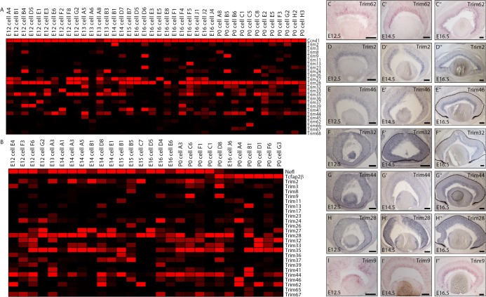
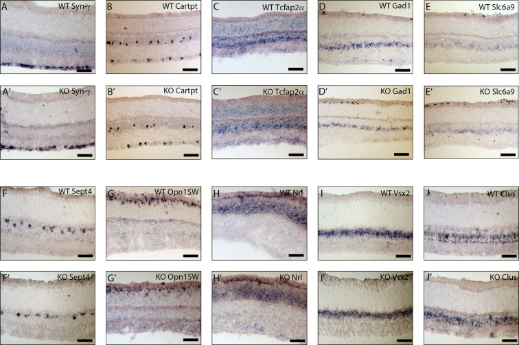
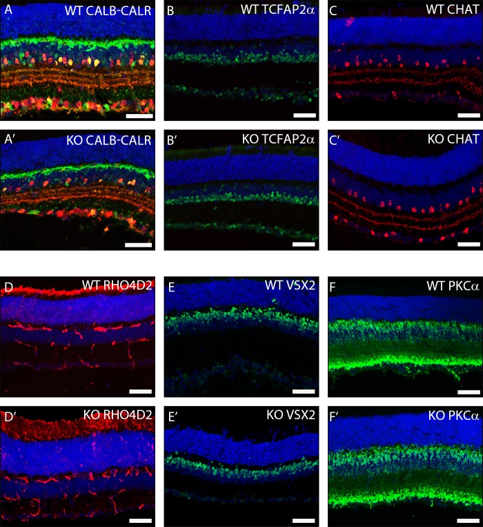
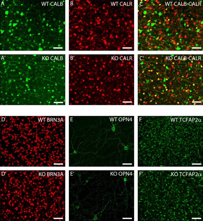
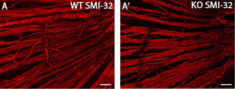
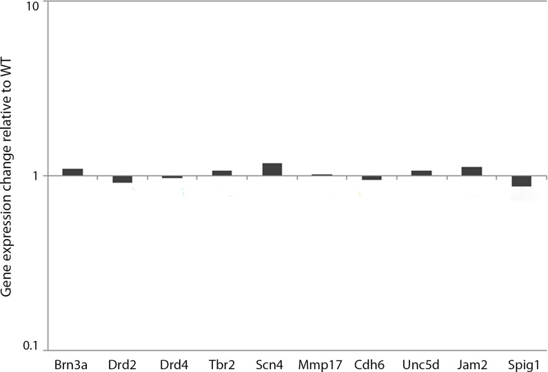
To better understand the mechanisms that govern the development of retinal neurons, it is critical to gain additional insight into the specific intrinsic factors that control cell fate decisions and neuronal maturation. In the developing mouse retina, Atoh7, a highly conserved transcription factor, is essential for retinal ganglion cell development. Moreover, Atoh7 expression in the developing retina occurs during a critical time period when progenitor cells are in the process of making cell fate decisions. We performed transcriptome profiling of Atoh7+ individual cells isolated from mouse retina. One of the genes that we found significantly correlated with Atoh7 in our transcriptomic data was the E3 ubiquitin ligase, Trim9. The correlation between Trim9 and Atoh7 coupled with the expression of Trim9 in the early mouse retina led us to hypothesize that this gene may play a role in the process of cell fate determination. To address the role of Trim9 in retinal development, we performed a functional analysis of Trim9 in the mouse and did not detect any morphological changes in the retina in the absence of Trim9. Thus, Trim9 alone does not appear to be involved in cell fate determination or early ganglion cell development in the mouse retina. We further hypothesize that the reason for this lack of phenotype may be compensation by one of the many additional TRIM family members we find expressed in the developing retina.
Conflict of interest statement
The authors have declared that no competing interests exist.
Figures
Similar articles
-
Reprogramming amacrine and photoreceptor progenitors into retinal ganglion cells by replacing Neurod1 with Atoh7.Development. 2013 Feb 1;140(3):541-51. doi: 10.1242/dev.085886. Development. 2013. PMID: 23293286 Free PMC article.
-
Transcriptome of Atoh7 retinal progenitor cells identifies new Atoh7-dependent regulatory genes for retinal ganglion cell formation.Dev Neurobiol. 2014 Nov;74(11):1123-40. doi: 10.1002/dneu.22188. Epub 2014 May 22. Dev Neurobiol. 2014. PMID: 24799426 Free PMC article.
-
TRIM9, a novel brain-specific E3 ubiquitin ligase, is repressed in the brain of Parkinson's disease and dementia with Lewy bodies.Neurobiol Dis. 2010 May;38(2):210-8. doi: 10.1016/j.nbd.2010.01.007. Epub 2010 Jan 18. Neurobiol Dis. 2010. PMID: 20085810 Free PMC article.
-
The TRIM9/TRIM67 neuronal interactome reveals novel activators of morphogenesis.Mol Biol Cell. 2021 Feb 15;32(4):314-330. doi: 10.1091/mbc.E20-10-0622. Epub 2020 Dec 30. Mol Biol Cell. 2021. PMID: 33378226 Free PMC article.
-
TRIM proteins in hepatocellular carcinoma.J Biomed Sci. 2022 Sep 13;29(1):69. doi: 10.1186/s12929-022-00854-7. J Biomed Sci. 2022. PMID: 36100865 Free PMC article. Review.
Cited by
-
Trim9 regulates the directional differentiation of retinal Müller cells to retinal ganglion cells.Zhong Nan Da Xue Xue Bao Yi Xue Ban. 2023 Oct 28;48(10):1561-1571. doi: 10.11817/j.issn.1672-7347.2023.230108. Zhong Nan Da Xue Xue Bao Yi Xue Ban. 2023. PMID: 38432885 Free PMC article. Chinese, English.
-
Association of the SNP rs112369934 near TRIM66 Gene with POAG Endophenotypes in African Americans.Genes (Basel). 2021 Sep 15;12(9):1420. doi: 10.3390/genes12091420. Genes (Basel). 2021. PMID: 34573402 Free PMC article.
-
A hybrid technique for measurement of intra/extracellular proteins.PLoS One. 2023 May 4;18(5):e0282948. doi: 10.1371/journal.pone.0282948. eCollection 2023. PLoS One. 2023. PMID: 37141290 Free PMC article.
-
TRIM9 promotes Müller cell-derived retinal stem cells to differentiate into retinal ganglion cells by regulating Atoh7.In Vitro Cell Dev Biol Anim. 2023 Sep;59(8):586-595. doi: 10.1007/s11626-023-00807-w. Epub 2023 Oct 4. In Vitro Cell Dev Biol Anim. 2023. PMID: 37792226
-
Identification of key genes and pathways for melanoma in the TRIM family.Cancer Med. 2020 Dec;9(23):8989-9005. doi: 10.1002/cam4.3545. Epub 2020 Oct 28. Cancer Med. 2020. PMID: 33118318 Free PMC article.
References
-
- Holt CE, Bertsch TW, Ellis HM, Harris WA. Cellular determination in the Xenopus retina is independent of lineage and birth date. Neuron. 1988. March;1(1):15–26. - PubMed
-
- Turner DL, Snyder EY, Cepko CL. Lineage-independent determination of cell type in the embryonic mouse retina. Neuron. 1990;4(6):833–45. - PubMed
Publication types
MeSH terms
Substances
LinkOut - more resources
Full Text Sources
Other Literature Sources
Molecular Biology Databases
