. 2018 Oct 17;140(41):13260-13271.
doi: 10.1021/jacs.8b06923. Epub 2018 Oct 8.

The Zinc Linchpin Motif in the DNA Repair Glycosylase MUTYH: Identifying the Zn2+ Ligands and Roles in Damage Recognition and Repair

Affiliations

The Zinc Linchpin Motif in the DNA Repair Glycosylase MUTYH: Identifying the Zn2+ Ligands and Roles in Damage Recognition and Repair

Nicole N Nuñez et al. J Am Chem Soc. .

Abstract

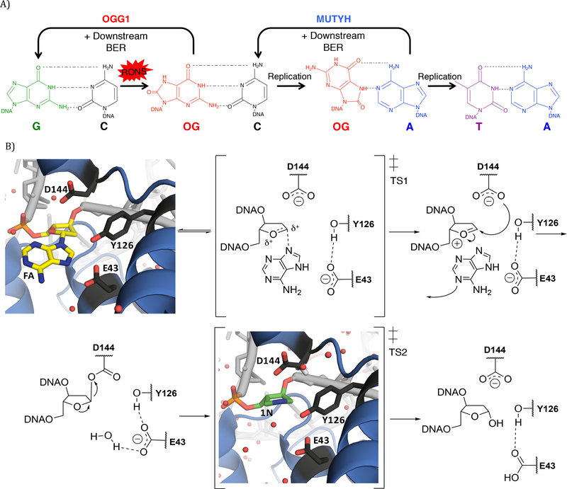
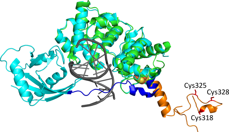
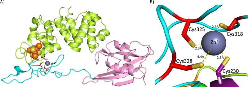
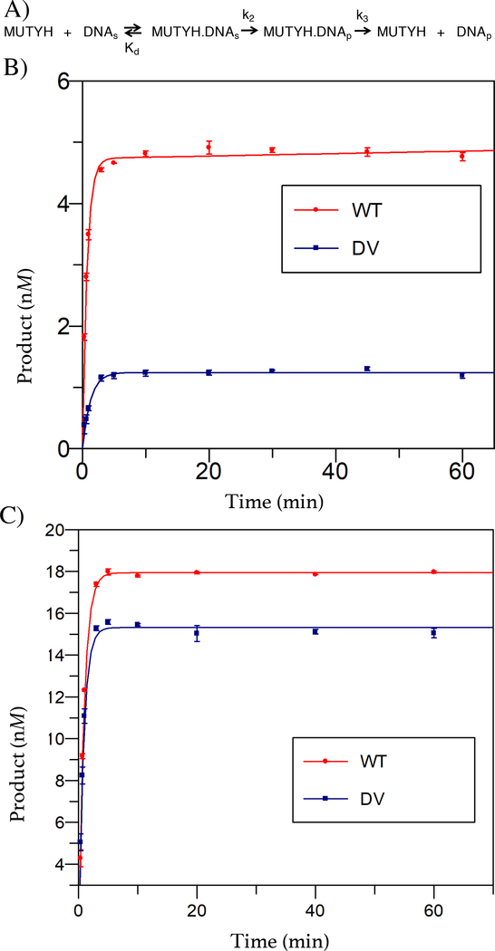
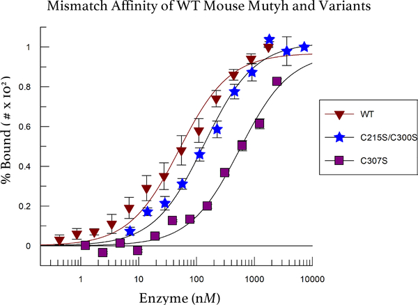
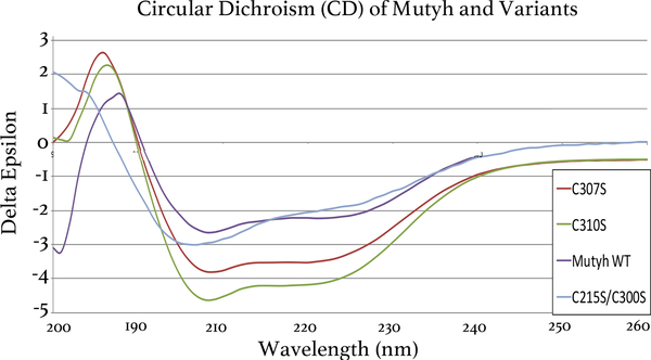
The DNA base excision repair (BER) glycosylase MUTYH prevents DNA mutations by catalyzing adenine (A) excision from inappropriately formed 8-oxoguanine (8-oxoG):A mismatches. The importance of this mutation suppression activity in tumor suppressor genes is underscored by the association of inherited variants of MUTYH with colorectal polyposis in a hereditary colorectal cancer syndrome known as MUTYH-associated polyposis, or MAP. Many of the MAP variants encompass amino acid changes that occur at positions surrounding the two-metal cofactor-binding sites of MUTYH. One of these cofactors, found in nearly all MUTYH orthologs, is a [4Fe-4S]2+ cluster coordinated by four Cys residues located in the N-terminal catalytic domain. We recently uncovered a second functionally relevant metal cofactor site present only in higher eukaryotic MUTYH orthologs: a Zn2+ ion coordinated by three Cys residues located within the extended interdomain connector (IDC) region of MUTYH that connects the N-terminal adenine excision and C-terminal 8-oxoG recognition domains. In this work, we identified a candidate for the fourth Zn2+ coordinating ligand using a combination of bioinformatics and computational modeling. In addition, using in vitro enzyme activity assays, fluorescence polarization DNA binding assays, circular dichroism spectroscopy, and cell-based rifampicin resistance assays, the functional impact of reduced Zn2+ chelation was evaluated. Taken together, these results illustrate the critical role that the "Zn2+ linchpin motif" plays in MUTYH repair activity by providing for proper engagement of the functional domains on the 8-oxoG:A mismatch required for base excision catalysis. The functional importance of the Zn2+ linchpin also suggests that adjacent MAP variants or exposure to environmental chemicals may compromise Zn2+ coordination, and ability of MUTYH to prevent disease.


Conflict of interest statement

The authors declare no competing financial interests.

Figures

Figure 1.
Figure 1.
Proposed mechanism for adenine removal from 8-oxoG:A mismatches by MUTYH and orthologs. A) Endogenous and exogenous sources of reactive oxygen and nitrogen species (RONS) can chemically react with the guanine (G, green) to form 8-oxoG (or OG, red). The presence of 8-oxoG in DNA can result mutations via 8-oxoG:A mismatches. The BER glycosylase OGG1 removes 8-oxoG from 8-oxoG:C pairs, while MUTYH removes the A from 8-oxoG:A mismatches. The G:C base pair is restored by subsequent repair by downstream BER enzymes. B) The mechanism of adenine removal from 8-oxoG:A mismatches by MUTYH is proposed to occur via two SN1 - like displacement steps resulting in the formation of an abasic (AP) site product. The A nucleotide that is targeted is depicted in the mechanism with the rest of the DNA polymer projecting from the 5’ or 3’ side indicated as “ODNA”. A crystal structure of Geobacillus stearothermophilus (Gs) MutY (blue) bound to DNA (light grey) containing a noncleavable substrate base analog arabino 2’-fluoro-2’-deoxyadenosine, FA (yellow) (PDB ID: 3G0Q) demonstrates the positioning of key residues involved in catalysis. Gs MutY bound to DNA containing an azasugar transition state mimic, 1N (green) (PDB ID: 5DPK) also displays these participating amino acids, and position of the proposed water nucleophile. Amino acid residues of MutY that are in close proximity to the FA/1N nucleotides are labeled and colored in dark grey. Water molecules are depicted as red spheres and elements are depicted with oxygen in red, nitrogen in blue, and phosphorous in orange.
Figure 2.
Figure 2.
N-terminal fragment crystal structure of Homo sapiens MUTYH (PDB ID: 3N5N, green). MUTYH residues 293 through 353 correspond to the IDC (orange) aligned to the ortholog structure of Geobacillus stearothermophilus MutY (PDB ID: 5DPK, light blue) bound to DNA (grey), demonstrating the significantly shorter IDC (residues 215 through 234, dark blue) found in prokaryotes. Image also depicts established Zn2+ ion chelating ligands Cys318, Cys325 and Cys328 (red). The Fe-S cluster is depicted as orange and yellow spheres.
Figure 3.
Figure 3.
QM/MM energy-minimized structure of human MUTYH highlighting the four Cys residues interactions with Zn2+. A) A representative structure of human MUTYH from an MD simulation in explicit solvent after QM/MM minimization of the Zn2+-binding site (see Methods). Color coding is as follows: C-terminal, pink; N-terminal, green; IDC, light blue; three previously identified Cys ligands (Cys318, Cys325 and Cys328 in human MUTYH; corresponds to Cys300, Cys307 and Cys310 in mouse Mutyh respectively), red; newly identified fourth Cys ligand (Cys230 in human MUTYH; corresponds to Cys215 in mouse Mutyh), purple; Cys residues coordinating the Fe-S cluster, orange, with the Fe-S cluster designated by orange (Fe) and yellow (S) spheres. B) Close up of MUTYH coordinating Zn2+ via four S atoms from Cys230, Cys318, Cys325 and Cys328 (waters removed for clarity). Notably, a single water molecule mediates the interaction between Cys328 and Zn2+ (Figure S5). Distances are drawn as yellow dash lines and elements with oxygen in red sticks; nitrogen, in blue; sulfur, in yellow. Cys residues 230, 318, 325 and 328 in human MUTYH correspond to 215, 300, 307 and 310 respectively in mouse Mutyh.
Figure 4.
Figure 4.
Adenine glycosylase activity of wild type (WT) and double variant (DV) Cys215Ser/Cys300Ser mouse Mutyh. (A) Minimal kinetic scheme used to evaluate binding (Kd) and subsequent removal of adenine from an 8-oxoG:A base pair-containing duplex substrates. The rate constant k2, reports on all steps involved in base excision to form an abasic site product. Release of the product from MUTYH is represented by the rate constant k3. (B) MTO glycosylase assays reveal a reduced active fraction, as evidence by the reduced amplitude of the “burst” phase for the double variant (DV) compared to wild type (WT) mouse Mutyh. (C) Graph of WT and DV Mutyh activity under single-turnover conditions ([active enzyme]>>[DNA]) to determine the intrinsic rate of glycosidic bond cleavage, k2. The data are shown with average and standard error bars from experiments performed with a minimum of three separate sample aliquots.
Figure 5.
Figure 5.
Mismatch Affinity of WT and Zn2+ Linchpin Mouse Mutyh Variants. Fluorescence polarization assays of WT mouse Mutyh and Zn2+ linchpin Cys to Ser variants that have reduced Zn2+ loading (40%, double variant Cys215Ser/Cys300Ser; 10%, Cys307Ser) show reduced affinity for an 8-oxoG:FA duplex. The Kd values for the double variant Cys215Ser/Cys300Ser and Cys307Ser variant are 136 ± 14 and 560 ± 80 nM, respectively, compared to WT Mutyh, 50 ± 8 nM. Additional binding data are shown in Figure S7. Cys215, Cys300 and Cys307 in mouse Mutyh correspond to Cys230, Cys318 and Cys325 in human MUTYH.
Figure 6.
Figure 6.
Circular Dichroism (CD) Spectra of WT MouseMutyh and Cys ligand variants. CD signal is normalized to the protein concentration by conversion of millidegrees (θ) to delta epsilon (Δε, M−1cm−1) and are plotted versus wavelength (nm). The CD demonstrates that the dramatic loss in percent activity for Zn2+ compromised variants (Cys215, Cys300, Cys307 and Cys310 in mouse Mutyh correspond to Cys230, Cys318, Cys325 and Cys328 in MUTYH, respectively) is not due to a complete loss of protein structure, rather Zn2+ coordination plays a local structural role (Figure S8).

References

    1. Banda DM, Nuñez NN, Burnside MA, Bradshaw KM and David SS (2017) Free Radical Biology and Medicine 107, 202–215. - PMC - PubMed
    1. David SS, O’Shea VL and Kundu S (2007) Nature 447 (7147), 941–950. - PMC - PubMed
    1. Al-Tassan N, Chmiel NH, Maynard J, Fleming N, Livingston AL, Williams GT, Hodges AK, Davies DR, David SS, Sampson JR and Cheadle JR (2002) Nat. Genet 30 (2), 227–232. - PubMed
    1. Wallace SS, Murphy DL and Sweasy JB (2012) Cancer Letters 327 (1–2), 73–89. - PMC - PubMed
    1. Mazzei F, Viel A and Bignami M (2013) Mutat. Res 743–744, 33–43. - PubMed

Publication types