. 2018 Sep 11;19(9):2707.
doi: 10.3390/ijms19092707.

Cellular Sources and Regional Variations in the Expression of the Neuroinflammatory Marker Translocator Protein (TSPO) in the Normal Brain

Affiliations

Cellular Sources and Regional Variations in the Expression of the Neuroinflammatory Marker Translocator Protein (TSPO) in the Normal Brain

Calina Betlazar et al. Int J Mol Sci. .

Abstract

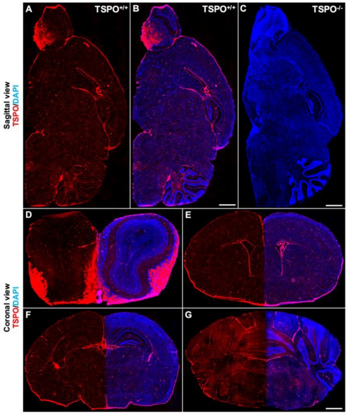
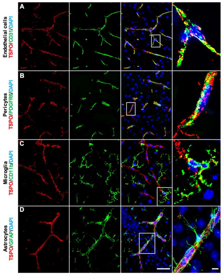
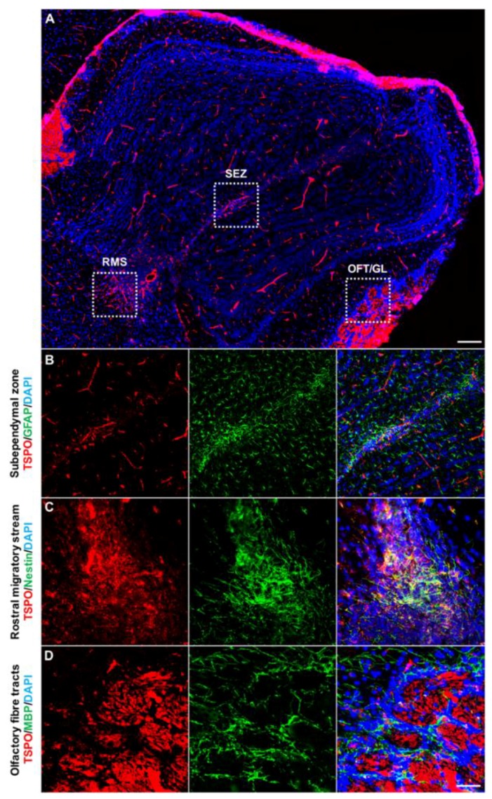
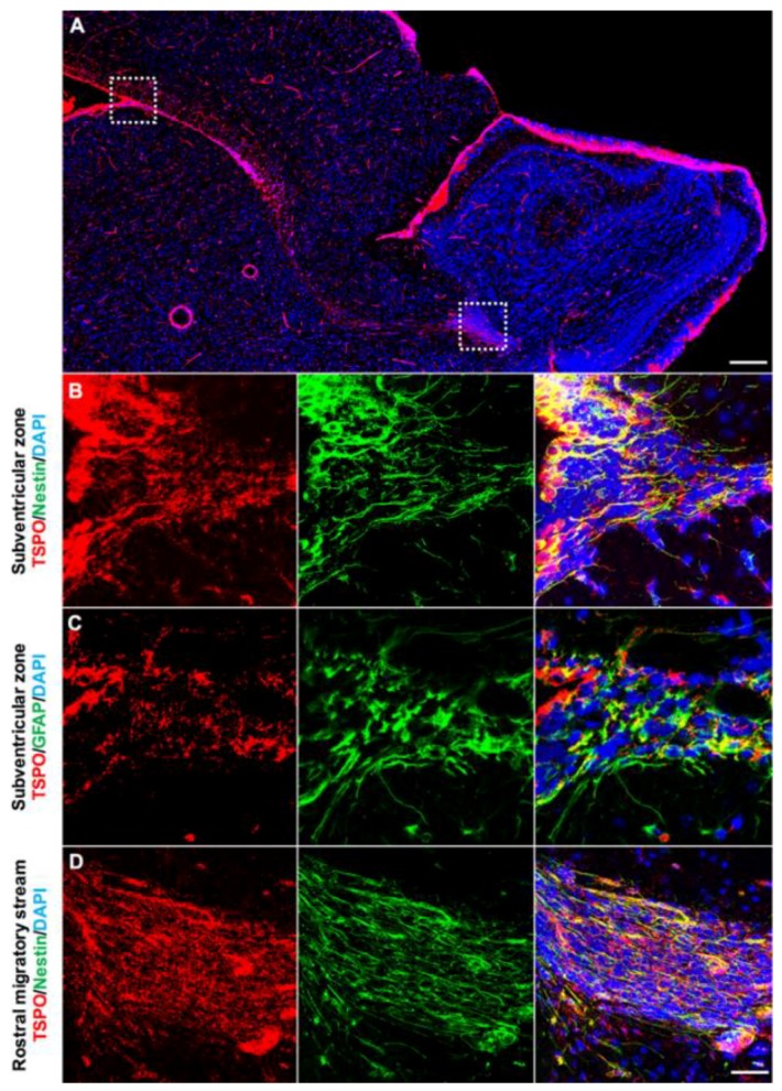
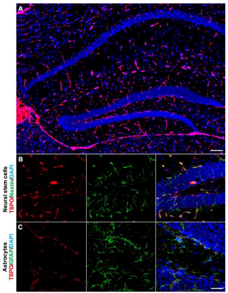
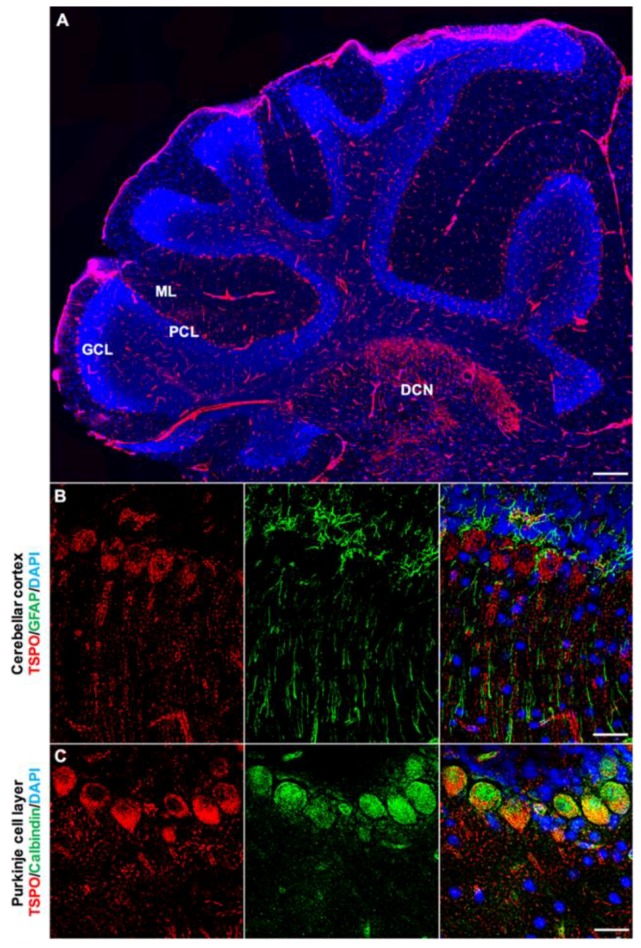
The inducible expression of the mitochondrial translocator protein 18 kDa (TSPO) by activated microglia is a prominent, regular feature of acute and chronic-progressive brain pathology. This expression is also the rationale for the continual development of new TSPO binding molecules for the diagnosis of "neuroinflammation" by molecular imaging. However, there is in the normal brain an ill-defined, low-level constitutive expression of TSPO. Taking advantage of healthy TSPO knockout mouse brain tissue to validate TSPO antibody specificity, this study uses immunohistochemistry to determine the regional distribution and cellular sources of TSPO in the normal mouse brain. Fluorescence microscopy revealed punctate TSPO immunostaining in vascular endothelial cells throughout the brain. In the olfactory nerve layers and glomeruli of the olfactory bulb, choroid plexus and ependymal layers, we confirm constitutive TSPO expression levels similar to peripheral organs, while some low TSPO expression is present in regions of known neurogenesis, as well as cerebellar Purkinje cells. The distributed-sparse expression of TSPO in endothelial mitochondria throughout the normal brain can be expected to give rise to a low baseline signal in TSPO molecular imaging studies. Finally, our study emphasises the need for valid and methodologically robust verification of the selectivity of TSPO ligands through the use of TSPO knockout tissues.

Keywords: immunohistochemistry; mitochondria; neuroinflammation; positron emission tomography; translocator protein.


Conflict of interest statement

The authors declare no conflict of interest.

Figures

Figure 1
Figure 1
Global immunofluorescence overview of TSPO (translocator protein 18 kDa) expression across major brain regions. (A) Sagittal section from a wildtype mouse (TSPO+/+) demonstrates widespread TSPO expression (red) across all brain regions using a specific antibody against TSPO (anti-PBR, ab109497). (B) Sagittal section from a wildtype mouse with TSPO expression in red and 4’,6-diamidino-2-phenylindole (DAPI) in blue highlighting structural features. Strong TSPO immunoreactivity is present in the olfactory bulb, the choroid plexus and the ependyma of the ventricular system, and cerebellum. (C) Sagittal section from a TSPO knockout mouse (TSPO−/−) confirming the absence of TSPO expression (DAPI in blue). (D) Coronal view of the olfactory bulb demonstrates strong TSPO expression in the olfactory nerve layers and glomeruli, and concentrated expression in the subependymal zone. (E) TSPO expression is present in the subventricular zone and the ependymal cells of the ventricles and choroid plexus. (F) Coronal view of the hippocampal region with discernible TSPO expression observed in the dentate gyrus. (G) Cerebellar/brainstem TSPO expression is strongly present in the molecular layer of the cerebellar cortex and fiber tracts of the brainstem. Scale bars = 500 µm.
Figure 2
Figure 2
TSPO expression in major cell types of the normal mouse brain. (A) Double immunofluorescence staining for TSPO (red) demonstrates punctate staining which colocalizes strongly with CD31+ vascular endothelial cells (green). This is observed in larger blood vessels, smaller arterioles and venules, as well as capillaries. Punctate staining is indicative of mitochondrial localization. (B) TSPO expression (red) and PDGFRβ+ pericytes/smooth muscle cells (green) colocalize on larger blood vessel walls. (C) No clearly discernible TSPO expression (red) is observed in CD11b+ microglia (green). (D) TSPO expression (red) is not observed in GFAP+ astrocytes (green). Perivascular astrocytic endfeet and processes surround CD31+ endothelial cells of blood vessels, where TSPO strictly colocalizes. Scale bars = 20 µm, 10 µm for box insets.
Figure 3
Figure 3
TSPO expression across the olfactory bulb in the normal mouse brain. (A) Sagittal view of TSPO expression (red) across the olfactory bulb. Structural features of the region highlighted with DAPI (blue). (B) Concentrated TSPO expression (red) is observed in the subependymal zone, which houses a neural stem cell niche derived from the rostral migratory stream (RMS). TSPO colocalizes at low levels with GFAP+ (green) cells. (C) TSPO is present at the base of the RMS, entering into the olfactory bulb. Here, TSPO strongly colocalizes with Nestin+ (green) cells. (D) TSPO expression (red) is also observed in the olfactory nerve layer and glomerular layer, though does not colocalize with MBP+ oligodendrocytes (green) of the region. Scale bars = 300 µm for (A), 20 µm for (BD). RMS = rostral migratory stream, SEZ = subependymal zone, OFT = olfactory fibre tracts, GL = glomerular layer.
Figure 4
Figure 4
TSPO is expressed in the neurogenic regions of the subventricular zone and the rostral migratory stream (RMS). (A) Strong TSPO immunofluorescence (red) is observed in the subventricular zone adjacent to the ependymal lining of the ventricles, and extends through the RMS to the neurogenic niche of the olfactory bulb. (B) TSPO expression (red) is observed in Nestin+ neural stem/progenitor cells (green) in the subventricular zone (sagittal view). (C) In the RMS, TSPO expression in Nestin+ progenitor cells (green) is also high (sagittal view). (D) TSPO expression (red) also colocalizes with GFAP in neural stem/progenitor cells (green) in the subventricular zone (coronal view), confirming the presence of TSPO in neural stem/progenitor cells. Scale bars = 300 µm for (A), 20 µm for (BD).
Figure 5
Figure 5
TSPO expression in the hippocampal region of the normal mouse brain. (A) TSPO expression (red) and DAPI (blue) to label the distinctive structural features of the region. Strong TSPO immunoreactivity is observed within the subgranular zone of the dentate gyrus and the ependyma. (B) TSPO (red) colocalizes at low levels with Nestin+ cell bodies (green), but not processes, in the subgranular zone of the dentate gyrus. (C) TSPO expression (red) does not colocalize with GFAP+ astrocytes (green) of the hippocampus, though is present in GFAP+ neural stem/progenitor cells of the subgranular zone, further confirming the expression of TSPO in neural stem/progenitor cells. Scale bar = 300 µm for (A), 20 µm for (B,C).
Figure 6
Figure 6
TSPO expression in the cerebellum of the normal mouse brain. (A) Sagittal view of TSPO expression (red) across the cerebellar region. Structural features of the region highlighted with DAPI (blue) to delineate cerebellar cortical layers. TSPO is expressed in the Purkinje cell layer and the molecular layer. Also of note is salient TSPO expression in the deep cerebellar nuclei. (B) Immunofluorescence staining with TSPO (red) and GFAP (green) in the Purkinje cell layer. TSPO is clearly discernible in Purkinje cells and the molecular layer, which contains dendritic projections. GFAP+ astrocytes/radial glia are also found in this region, though do not express TSPO. (C) Calbindin (green) and TSPO (red) immunostaining confirms significant colocalization of TSPO in the perinuclear region of Purkinje cells. Scale bars = 300 µm for (A), 20 µm for (B,C). ML = molecular layer, PCL = Purkinje cell layer, GCL = granule cell layer, DCN = deep cerebellar nuclei.

References

    1. Papadopoulos V., Baraldi M., Guilarte T.R., Knudsen T.B., Lacapere J.J., Lindemann P., Norenberg M.D., Nutt D., Weizman A., Zhang M.R., et al. Translocator protein (18kda): New nomenclature for the peripheral-type benzodiazepine receptor based on its structure and molecular function. Trends Pharmacol. Sci. 2006;27:402–409. doi: 10.1016/j.tips.2006.06.005. - DOI - PubMed
    1. Liu G.J., Middleton R.J., Hatty C.R., Kam W.W., Chan R., Pham T., Harrison-Brown M., Dodson E., Veale K., Banati R.B. The 18 kda translocator protein, microglia and neuroinflammation. Brain Pathol. 2014;24:631–653. doi: 10.1111/bpa.12196. - DOI - PMC - PubMed
    1. Banati R.B., Middleton R.J., Chan R., Hatty C.R., Kam W.W., Quin C., Graeber M.B., Parmar A., Zahra D., Callaghan P., et al. Positron emission tomography and functional characterization of a complete pbr/tspo knockout. Nat. Commun. 2014;5:5452. doi: 10.1038/ncomms6452. - DOI - PMC - PubMed
    1. Gut P., Zweckstetter M., Banati R.B. Lost in translocation: The functions of the 18-kd translocator protein. Trends Endocrinol. Metab. 2015;26:349–356. doi: 10.1016/j.tem.2015.04.001. - DOI - PMC - PubMed
    1. Liu G.J., Middleton R.J., Kam W.W., Chin D.Y., Hatty C.R., Chan R.H., Banati R.B. Functional gains in energy and cell metabolism after tspo gene insertion. Cell Cycle. 2017;16:436–447. doi: 10.1080/15384101.2017.1281477. - DOI - PMC - PubMed

LinkOut - more resources