. 2018 Oct 31;132(4):jcs221556.
doi: 10.1242/jcs.221556.

Claudin-4 reconstituted in unilamellar vesicles is sufficient to form tight interfaces that partition membrane proteins

Affiliations

Claudin-4 reconstituted in unilamellar vesicles is sufficient to form tight interfaces that partition membrane proteins

Brian Belardi et al. J Cell Sci. .

Abstract

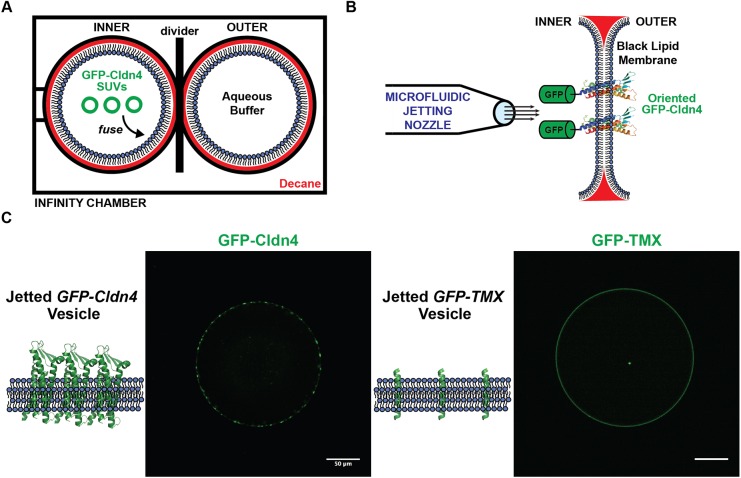
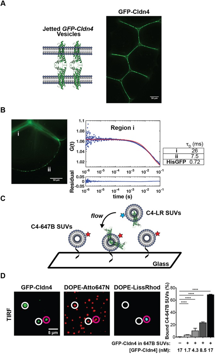
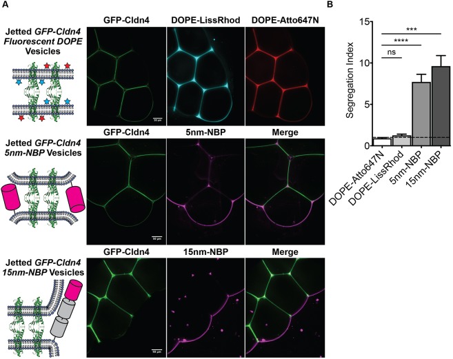
Tight junctions have been hypothesized to act as molecular fences in the plasma membrane of epithelial cells, helping to form differentiated apical and basolateral domains. While this fence function is believed to arise from the interaction of four-pass transmembrane claudins, the complexity of tight junctions has made direct evidence of their role as a putative diffusion barrier difficult to obtain. Here, we address this challenge by reconstituting claudin-4 into giant unilamellar vesicles using microfluidic jetting. We find that reconstituted claudin-4 alone can form adhesive membrane interfaces without the accessory proteins that are present in vivo By controlling the molecular composition of the inner and outer leaflets of jetted vesicle membranes, we show that claudin-4-mediated interfaces can drive partitioning of extracellular membrane proteins with ectodomains as small as 5 nm but not of inner or outer leaflet lipids. Our findings indicate that homotypic interactions of claudins and their small size can contribute to the polarization of epithelial cells.

Keywords: Cell adhesion; Epithelial cells; Polarity; Reconstitution; Tight junction; Transmembrane protein.


Conflict of interest statement

Competing interestsThe authors declare no competing or financial interests.

Figures

Fig. 1.
Fig. 1.
Reconstitution of GFP–Cldn4 in jetted vesicles. (A) Infinity chamber configuration. (B) GFP–Cldn4 black lipid membrane scheme. (C) Fluorescence micrographs of single jetted vesicles containing either GFP–Cldn4 (left) or GFP–TMX (right). Scale bars: 50 μm.
Fig. 2.
Fig. 2.
Cldn4 is sufficient to form membrane interfaces. (A) Diagram and fluorescence micrograph of jetted GUV assemblies containing GFP–Cldn4. (B) FCS autocorrelation curves were constructed for GFP–Cldn4 at interfaces, denoted region i, and at free membrane regions, denoted region ii (left). Diffusion times (right, mean, n=6 for all conditions) were then calculated for the different regions. (C) Scheme for the single SUV binding assay. (D) TIRF micrographs and quantification of the single SUV binding assay. The vesicles highlighted by magenta and white circles are bound and unbound SUVs, respectively. Data are presented as mean±s.d. (n=13, 9, 13, 12 and 10, respectively with n>500 particles counted per sample). ****P<0.0001 (two-tailed unpaired t-test). Scale bars: 50 μm (A,B), 5 μm (D).
Fig. 3.
Fig. 3.
Cldn4–Cldn4 interfaces drive the segregation of membrane-bound proteins. (A) Diagrams and fluorescent micrographs of jetted GFP–Cldn4 GUVs containing fluorescent lipids (upper), fluorescent 5 nm NBPs (middle) or fluorescent 15 nm NBPs (bottom). (B) Quantification of extent of segregation for inner leaflet lipids, DOPE-Atto647N, outer leaflet lipids, DOPE-LissRhod, and 5 nm NBP and 15 nm NBPs. Segregation Index=[Fluorescence Intensityfree membrane/(Fluorescence Intensityinterface/2)]. Data are presented as mean±s.d. (n=5 for all conditions). ***P<0.001; ****P<0.0001; ns, not significant (two-tailed unpaired t-test). Scale bars: 50 μm.

References

    1. Baas A. F., Kuipers J., van der Wel N. N., Batlle E., Koerten H. K., Peters P. J. and Clevers H. C. (2004). Complete polarization of single intestinal epithelial cells upon activation of LKB1 by STRAD. Cell 116, 457-466. 10.1016/S0092-8674(04)00114-X - DOI - PubMed
    1. Bacia K., Haustein E. and Schwille P. (2014). Fluorescence correlation spectroscopy: principles and applications. Cold Spring Harb. Protoc. 2014, 709-725. 10.1101/pdb.top081802 - DOI - PubMed
    1. Cole C. M., Brea R. J., Kim Y. H., Hardy M. D., Yang J. and Devaraj N. K. (2015). Spontaneous reconstitution of functional transmembrane proteins during bioorthogonal phospholipid membrane synthesis. Angew. Chem. Int. Ed. 54, 12738-12742. 10.1002/anie.201504339 - DOI - PMC - PubMed
    1. Dezi M., Cicco A. D., Bassereau P. and Lévy D. (2013). Detergent-mediated incorporation of transmembrane proteins in giant unilamellar vesicles with controlled physiological contents. Proc. Natl. Acad. Sci. 110, 7276-7281. 10.1073/pnas.1303857110 - DOI - PMC - PubMed
    1. Diamond J. M. (1977). Twenty-first Bowditch lecture. The epithelial junction: bridge, gate, and fence. Physiologist 20, 10-18. - PubMed

Publication types

LinkOut - more resources