. 2018 Oct 15;201(8):2354-2368.
doi: 10.4049/jimmunol.1800671. Epub 2018 Sep 12.

Influenza A Virus Infection Causes Chronic Lung Disease Linked to Sites of Active Viral RNA Remnants

Affiliations

Influenza A Virus Infection Causes Chronic Lung Disease Linked to Sites of Active Viral RNA Remnants

Shamus P Keeler et al. J Immunol. .

Abstract

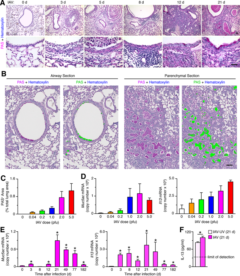
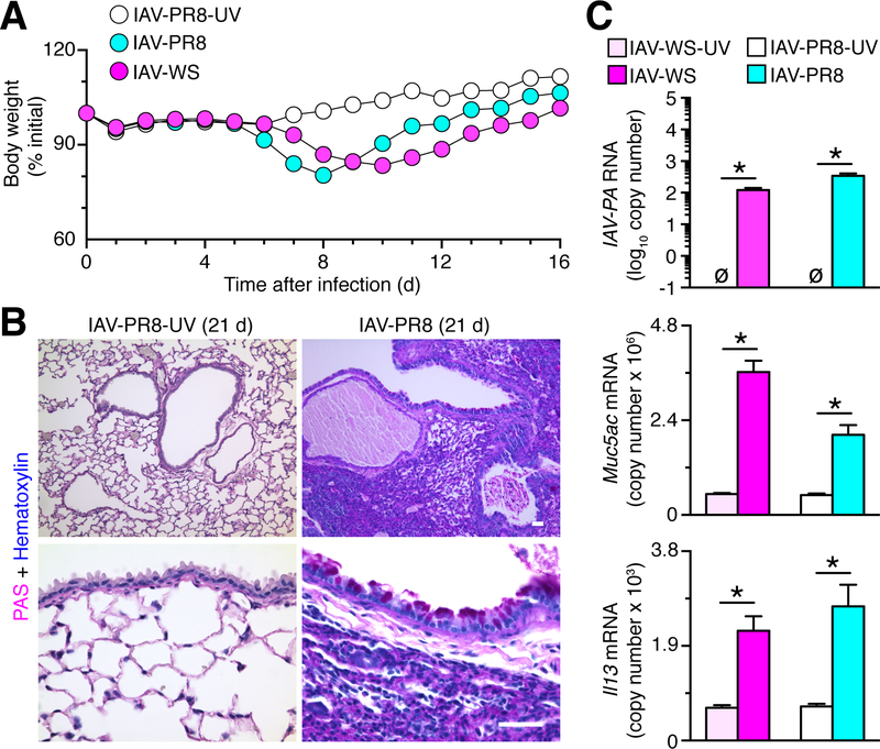
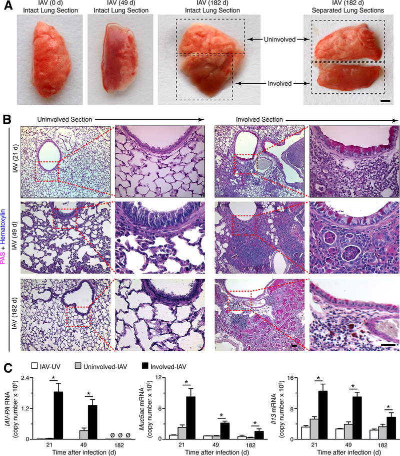
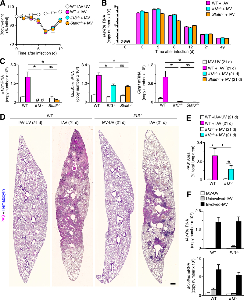
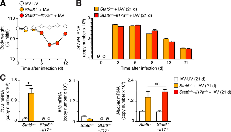
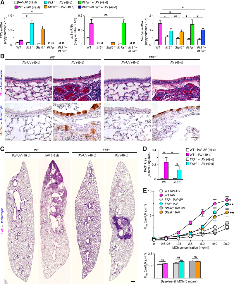
Clinical and experimental observations suggest that chronic lung disease is linked to respiratory viral infection. However, the long-term aspect of this relationship is not yet defined using a virus that replicates at properly high levels in humans and a corresponding animal model. In this study, we show that influenza A virus infection achieves 1 × 10⁶-fold increases in viral load in the lung and dose-dependent severity of acute illness in mice. Moreover, these events are followed by persistence of negative- and positive-strand viral RNA remnants for 15 wk and chronic lung disease for at least 26 wk postinfection. The disease is manifested by focal areas of bronchiolization and mucus production that contain increased levels of viral RNA remnants along with mucin Muc5ac and Il13 mRNA compared with uninvolved areas of the lung. Excess mucus production and associated airway hyperreactivity (but not fibrosis or emphysema) are partially attenuated with loss of IL-13 production or signaling (using mice with IL-13 or STAT6 deficiency). These deficiencies cause reciprocal increases in l17a mRNA and neutrophils in the lung; however, none of these disease endpoints are changed with IL-13/IL-17a compared with IL-13 deficiency or STAT6/IL-17a compared with STAT6 deficiency. The results establish the capacity of a potent human respiratory virus to produce chronic lung disease focally at sites of active viral RNA remnants, likely reflecting locations of viral replication that reprogram the region. Viral dose dependency of disease also implicates high-level viral replication and severity of acute infection as determinants of chronic lung diseases such as asthma and COPD with IL-13-dependent and IL-13/IL-17-independent mechanisms.


Conflict of interest statement

Disclosures

The authors have no financial conflicts of interest.

Figures

FIGURE 1.
FIGURE 1.
Acute illness leaves viral remnants after IAV infection. (A) Time course of body weight change in wild-type C56BL/6J mice after infection with IAV (WS/33 strain, 0–5 pfu). Mice were euthanized at body weight <75% of initial value. (B) Corresponding lung levels of IAV monitored with plaque-forming assay for conditions in (A) at 3 d after infection. (C) Corresponding lung levels of IAV RNA encoding the PA gene monitored with PCR assay for conditions in (B). (D) Time course of lung levels of IAV monitored with plaque-forming assay after infection with IAV (2 pfu). (E) Time course for IAV immunostaining in lung sections after infection with IAV (2 pfu). Bar=400 μm. (F) Time course for lung levels of IAV RNA after infection with IAV (2 pfu). (G) Corresponding IAV-PA RNA levels in tracheobronchial lymph nodes, spleen, and thymus. For (H), * indicates p<0.05 versus no infection control. (H) Scheme for strand-specific qPCR assay in which the negative (–) RNA strand of viral RNA (vRNA) (labeled in magenta) or the positive (+) RNA strand of viral RNA (cRNA, mRNA) (labeled in blue) were reverse transcribed to corresponding cDNA using tagged primers that included a portion complementary to each strand (green) and another portion that was not related to viral sequence (orange). The resulting tagged cDNA was amplified using PCR in which one primer was specific to the tagged portion of cDNA (orange) and another primer (green) and probe (black) were specific to viral sequence. (I) Validation of strand-specific tag-PCR assay in which (+) and (–) strand RNA standards were transcribed from a plasmid encoding the IAV-PA gene and cDNA was synthesized using primers specific for the (–) or (+) strand of viral RNA. (J) Time course of lung levels of IAV-PA negative- and positive-strand RNA detected using tag-PCR assay. (K) Time course of lung levels of IAV RNA encoding the NP gene (IAV-NP) detected using PCR assay. (L) Time course of lung levels of IAV (+) strand RNA encoding IAV-NP detected using tag-PCR assay. For (A)-(G) and (I)-(L), values are representative of 3 separate experiments (n=8 mice per condition in each experiment). Values for 0 d were not significantly different than UV-inactivated IAV (IAV-UV) at 3–182 d (data not shown).
FIGURE 2
FIGURE 2
Viral dose drives the development of chronic lung disease after IAV infection. (A) Time course for PAS-hematoxylin staining in lung sections after infection with IAV (WS/33 strain, 2 pfu). Bars=400 μm. (B) PAS-hematoxylin staining of lung sections for airway and parenchymal tissue at 21 d after IAV infection (2 pfu) with green colorization indicating computer-assigned PAS+ areas. Bar = 200 μm. (C) Quantitation of PAS+ area based on computer-assignment and quantification for conditions in (B). (D) Lung levels of Muc5ac and Il13 mRNA at 21 d after infection with IAV (0–5 pfu). (E) Time course for lung levels of Muc5ac and Il13 mRNA after infection with IAV (2 pfu). (F) Levels of IL-13 protein in lung tissue at 21 d after infection with IAV or IAV-UV. Dashed lines indicate the lower limit of detection for the assay. For (A)-(F), values are representative of 3 separate experiments (n≥8 mice per condition in each experiment). * indicates p<0.05 versus no infection or IAV-UV control (no difference was found for no infection versus IAV-UV).
FIGURE 3.
FIGURE 3.
Severe acute illness leads to chronic lung disease after IAV-PR8 infection. (A) Time course of body weight after infection with IAV (PR8 strain, 10 pfu or WS/33 strain, 2 pfu) or corresponding UV-inactivated IAV-PR8 in wild-type C56BL/6J mice. (B) PAS and-hematoxylin staining of lung sections at 21 d after infection with IAV-PR8 (10 pfu) or IAV-PR8-UV. Bar=400 μm. (C) Corresponding lung levels of IAV-PA RNA and Muc5ac and Il13 mRNA for conditions in (B). For (A,C), values are representative of 3 separate experiments (n≥8 mice per condition in each experiment). For (C), * indicates p<0.05.
FIGURE 4.
FIGURE 4.
Identification of focal areas of chronic lung disease after IAV infection. (A) Representative photographs of lung samples with grossly uninvolved or involved sections at 49 and 182 d (without and with separation by dissection) after infection with IAV (WS33 strain, 2 pfu). Bar=1 mm. (B) Representative PAS-hematoxylin staining of uninvolved or involved lung sections at 21, 49, and 182 d after infection with IAV (2 pfu). Bars=400 μm. (C) Levels of IAV-PA RNA and Muc5ac and Il13 mRNA in lung samples for conditions in (B) and control IAV-UV. For (A)-(C), values are representative of 3 separate experiments (n≥8 mice per condition in each experiment). For (C), * indicates p<0.05 versus uninvolved section.
FIGURE 5.
FIGURE 5.
IL-13 induction and STAT6 activation are linked to chronic lung disease after IAV infection. (A) Time course of body weight change in wild-type (WT), Il13–/–, and Stat6–/– mice on the indicated days after infection with IAV (2 pfu) or IAV-UV. (B) Time course for lung levels of IAV-PA RNA in the same mouse strains after infection with IAV (2 pfu). (C) Lung levels of Il13, Muc5ac, and Clca1 mRNA in the same mouse strains at 21 d after infection with IAV (2 pfu) or IA-UV. (D) PAS-hematoxylin staining of lung sections from WT and Il13–/–mice at 21 d after infection with IAV (2 pfu) or IAV-UV. Bar=1 mm. (E) Quantitation of PAS-positive areas in images in (D). (F) Quantitation of PAS+ area for conditions in (D). (F) Levels of IAV-PA RNA and Muc5ac mRNA in uninvolved or involved sections of lung from WT and Il13–/– mice at 21 d after infection with IAV or IAV-UV. For (A-C) and (E-F), values are representative of 3 separate experiments (n≥8 mice per condition in each experiment). * indicates p<0.05; ns=nonsignificant.
FIGURE 6.
FIGURE 6.
Reciprocal IL-17a induction after IL-13 blockade is not linked to chronic lung disease after IAV infection. (A) Lung levels of Il17a and Il17f mRNA in WT, Il13–/–, Il17a–/–, and Il13–/–Il17a–/– mice at 21 d after infection with IAV (2 pfu) or IA-UV. (B) Time course of body weight change in the same mouse strains as (A) after infection with IAV (2 pfu) or IAV-UV. (C) Time course for lung levels of IAV-PA RNA for conditions in (B). (D) Lung levels of Il13, Muc5ac, and Clca1 mRNA in same mouse strains as (A) at 21 d after infection with IAV (2 pfu) or IAV-UV. (E) Levels of indicated immune cells in BAL fluid for conditions in (D). (F) Levels of airway reactivity using response of respiratory system resistance (RRS) to inhaled methacholine (MCh) and for baseline RRS for conditions in (D). For (A-F), values are representative of 3 separate experiments (n=5–8 mice per condition in each experiment). For (A-E), * indicates p<0.05; for (F), * indicates p<0.05 versus IAV-UV and ** indicates p<0.05 versus corresponding IAV-UV and IAV conditions.
FIGURE 7.
FIGURE 7.
Reciprocal IL-17a induction due to STAT6-deficiency is not linked to chronic lung disease after IAV infection. (A) Time course of body weight change on the indicated days in Stat6–/– and Stat6–/––Il17a–/– mice after infection with IAV (2 pfu) or IAV-UV. (B) Time course for lung levels of IAV-PA RNA in same mouse strains as (A) after infection with IAV (2 pfu). (C) Lung levels of Il17a, Il13, and Muc5ac mRNA in same mouse strains as (A) at 21 d after infection with IAV (2 pfu) or IA-UV. For (A), (B), (C), values are representative of 3 separate experiments (n≥5 mice per condition in each experiment). * indicates p<0.05.
FIGURE 8.
FIGURE 8.
Persistence of IL-13–STAT6-linked chronic lung disease after IAV infection. (A) Lung levels of Il17a, Il13, and Muc5ac mRNA in WT, Il13–/–, Stat6–/–, Il17a–/–, and Il13–/–Il17a–/– mice at 49 d after infection with IAV (2 pfu) or IA-UV. (B) Representative PAS-hematoxylin staining and Muc5ac-hematoxylin immunostaining of lung sections from WT and Il13–/–mice at 49 d after infection with IAV (2 pfu) or IAV-UV. Bars=400 μm. (C) PAS-hematoxylin staining of lung sections from WT and Il13–/–mice at 21 d after infection with IAV (2 pfu) or IAV-UV. Bar=1 mm. (D) Quantitation of PAS-positive areas in images in (D). (E) Levels of airway reactivity using response RRS to inhaled MCh and for baseline RRS for conditions in (A). For (A)-(E), values are representative of 3 separate experiments (n≥8 mice per condition in each experiment). For (A), (C), and (D), * indicates p<0.05.
FIGURE 9.
FIGURE 9.
Persistent peribronchial and parenchymal fibrosis and pulmonary emphysema after IAV infection. (A) Gomori trichrome staining of lung sections from WT mice at 21 d after infection with IAV (2 pfu) or IAV-UV (WS/33 strain). Bar=1 mm. (B) Quantitation of trichrome-blue-positive areas for lung sections from the indicated mouse strains after infection with IAV or IAV-UV as described in (A). (C) Quantitation of trichrome-blue-positive areas for lung sections from the indicated mouse strains at 21 d after infection with IAV (10 pfu) or IAV-UV (PR8 strain). (D) Mean linear intercept values for indicated mouse strains after infection with IAV or IAV-UV (WS/33 strain) as described in (A). (E) Mean linear intercept values for indicated mouse strains after infection with IAV or IAV-UV (PR8 strain) as described in (C). For (B)-(E), values are representative of 3 separate experiments (n≥8 mice per condition in each experiment), and * indicates p<0.05.

References

    1. Holtzman MJ 2012. Asthma as a chronic disease of the innate and adaptive immune systems responding to viruses and allergens. J. Clin. Invest 122: 2741–2748. - PMC - PubMed
    1. Sigurs N, Gustafsson PM, Bjarnason R, Lundbeg F, Schmidt S, Sigurbergsson F, and Kjellman B. 2005. Severe respiratory syncytial virus bronchiolitis in infancy and asthma and allergy at age 13. Am. J. Respir. Crit. Care Med 171: 137–141. - PubMed
    1. Castro M, Schweiger T, Yin-Declue H, Ramkumar T, Christie C, Zheng J, Cohen R, Schechtman K, Strunk R, and Bacharier L. 2008. Cytokine response after severe respiratory syncytial virus bronchiolitis in early life. J. Allergy Clin. Immunol 122: 725–733. - PMC - PubMed
    1. Guilbert TW, Singh AM, Danov Z, Evans MD, Jackson DJ, Burton R, Roberg KA, Anderson EL, Pappas TE, Gangnon R, Gern JE, and Lemanske RF Jr. 2011. Decreased lung function after preschool wheezing rhinovirus illness in children at risk to develop asthma. J. Allergy Clin. Immunol 128: 532–538. - PMC - PubMed
    1. Kan-o K, Ramirez R, MacDonald MI, Rolph M, Rudd PA, Spann KM, Mahalingam S, Bardin PG, and Thomas BJ. 2017. Human metapneumovirus infection in chronic obstructive pulmonary disease: impact of glucocorticosteroids and inteferon. J. Inf. Dis 215: 1536–1545. - PubMed

Publication types

MeSH terms