. 2018 Sep 25;115(39):E9211-E9219.
doi: 10.1073/pnas.1810229115. Epub 2018 Sep 12.

Homologous recombination is an intrinsic defense against antiviral RNA interference

Affiliations

Homologous recombination is an intrinsic defense against antiviral RNA interference

Lauren C Aguado et al. Proc Natl Acad Sci U S A. .

Abstract

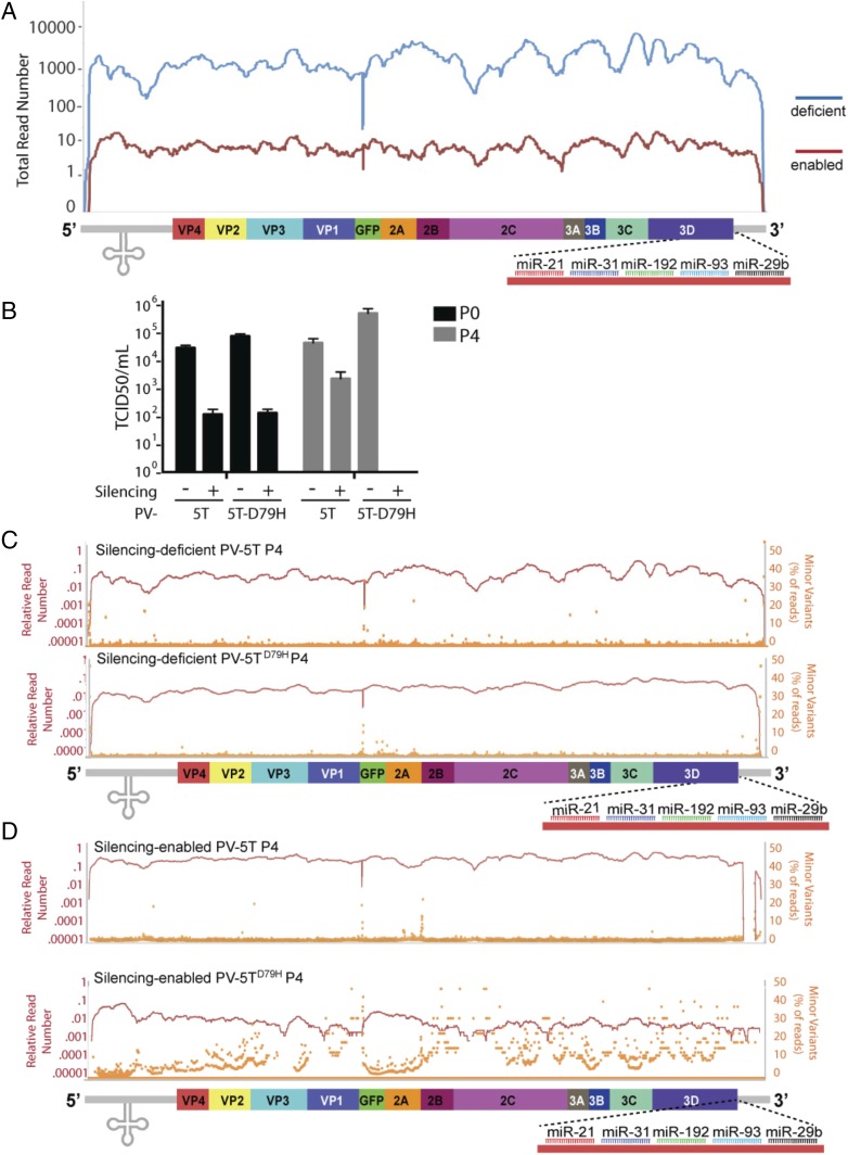
RNA interference (RNAi) is the major antiviral defense mechanism of plants and invertebrates, rendering the capacity to evade it a defining factor in shaping the viral landscape. Here we sought to determine whether different virus replication strategies provided any inherent capacity to evade RNAi in the absence of an antagonist. Through the exploitation of host microRNAs, we recreated an RNAi-like environment in vertebrates and directly compared the capacity of positive- and negative-stranded RNA viruses to cope with this selective pressure. Applying this defense against four distinct viral families revealed that the capacity to undergo homologous recombination was the defining attribute that enabled evasion of this defense. Independent of gene expression strategy, positive-stranded RNA viruses that could undergo strand switching rapidly excised genomic material, while negative-stranded viruses were effectively targeted and cleared upon RNAi-based selection. These data suggest a dynamic relationship between host antiviral defenses and the biology of virus replication in shaping pathogen prevalence.

Keywords: RNAi; homologous recombination; miRNA; virus adaptation; virus polarity.


Conflict of interest statement

The authors declare no conflict of interest.

Figures

Fig. 1.
Fig. 1.
The 5T cassette only restricts through miRNA machinery. (A) Diagrams of recombinant SeV-eGFP-5T and SeV-eGFP-5R genomes. The 5T (depicted with miRNA binding) and 5R (resistant to miRNA binding) cassettes are downstream of the GFP ORF, which is inserted between nucleoprotein and phosphoprotein ORFs. (B) Immunofluorescence microscopy of SeV-GFP-5T and -5R–infected MEFs. WT MEFS or Dicer (Dcr), Argonaute 2, or Argonaute 1, 3, and 4 knockout MEFs were infected at a MOI of 1 for 24 h. Cells were fixed and stained for SeV HN protein and DAPI. (C) Flow cytometry analysis of MEFs infected as stated in B. (D) FACS analysis of CRISPR KO library infected with SeV-GFP-5R or SeV-GFP-5T. Cells gated on FITC expression. (E) MAGeCK analysis of GFP+ CRISPR KO A549 cells infected with SeV-GFP-5T (D).
Fig. 2.
Fig. 2.
SeV-5T lacks the capacity to adapt to a silencing-enabled environment. (A) Diagram of recombinant SeV-5T where the 5T cassette is downstream of the nucleoprotein (N) ORF and the ORF encoding GFP is inserted between N and the phosphoprotein (P). All other viral proteins and miRNA targets are also depicted. (B) Western blot of whole-cell extracts from silencing-deficient cells infected with either SeV-5T or SeV (MOI = 0.01) at 12, 24, 36, and 48 hpi. Immunoblots depict the levels of N and GAPDH. (C) Western blot of whole-cell extracts as in B from silencing-enabled cells infected with either SeV-5T or SeV-(MOI = 0.01) at 12, 24, 36, and 48 hpi. (D) Infectious virus released by silencing-deficient and silencing-enabled cells infected with SeV-GFP-5T or 5R (MOI 1) after passage 1 (black) and passage 4 (gray). (E and F) Relative read numbers (red) and minor variants (orange) plotted along the SeV-5R genome from silencing-deficient cells (E) or silencing-enabled (F) cells at 48 hpi. (G and H) Relative read numbers (red) and minor variants (orange) plotted along the SeV-5T genome from silencing-deficient (G) or silencing-enabled (H) cells at 48 hpi.
Fig. 3.
Fig. 3.
SINV-5T rapidly adapts to RNAi-mediated silencing. (A) Diagrams of wild-type SINV (Top) and recombinant SINV-5T/-5R (Bottom), where the 5T cassette is downstream of the nonstructural ORF. (B) Western blot of whole-cell extracts from silencing-deficient cells infected with either SINV-5T or SINV-5R (MOI = 0.1) at 6, 12, 24, and 36 hpi. Immunoblots depict protein levels for SINV capsid (C or CP) and GAPDH. (C) Western blot as in B from silencing-enabled cells infected with either SINV-5T or SINV-5R (MOI = 0.1) at 6, 12, 24, and 36 hpi. (D) Relative read numbers (red) and minor variants (orange) from SINV-5R at 24 hpi (P0) and (P4) at 96 hpi in silencing-enabled cells. x axis denotes genomic alignment position. (E) Same as D from SINV-5T–infected silencing-enabled cells.
Fig. 4.
Fig. 4.
Positive-stranded RNA virus escape from RNAi-like pressure requires genome recombination. (A) Graph depicts the total number of reads mapping to each position along the viral genome for PV-5T after passage 0 in silencing-deficient (blue) and silencing-enabled (red) cells. Diagram of recombinant PV-5T where the 5T cassette is downstream of the PV ORF. All viral proteins and miRNA targets are depicted. (B) Infectious virus release of PV-5T WT or D79H after passage 0 (black) and passage 4 (gray) measured by TCID50 per milliliter. (C and D) Relative read numbers (red) and minor variants (orange) of PV5T-WT or PV-5T-D79H plotted along the respective viral genome after passage 4 in silencing-deficient cells (C) and silencing-enabled cells (D).

References

    1. Marraffini LA. CRISPR-Cas immunity in prokaryotes. Nature. 2015;526:55–61. - PubMed
    1. Doron S, et al. Systematic discovery of antiphage defense systems in the microbial pangenome. Science. 2018;359:eaar4120. - PMC - PubMed
    1. Koonin EV, Dolja VV, Krupovic M. Origins and evolution of viruses of eukaryotes: The ultimate modularity. Virology. 2015;479–480:2–25. - PMC - PubMed
    1. Prangishvili D, et al. The enigmatic archaeal virosphere. Nat Rev Microbiol. 2017;15:724–739. - PubMed
    1. tenOever BR. The evolution of antiviral defense systems. Cell Host Microbe. 2016;19:142–149. - PubMed

Publication types

Substances