. 2018 Sep 12;7(1):154.
doi: 10.1038/s41426-018-0155-5.

Genomic characterization and infectivity of a novel SARS-like coronavirus in Chinese bats

Affiliations

Genomic characterization and infectivity of a novel SARS-like coronavirus in Chinese bats

Dan Hu et al. Emerg Microbes Infect. .

Erratum in

  • Correction.
    [No authors listed] [No authors listed] Emerg Microbes Infect. 2020 Dec;9(1):2727. doi: 10.1080/22221751.2020.1857048. Emerg Microbes Infect. 2020. PMID: 33356975 Free PMC article. No abstract available.

Abstract

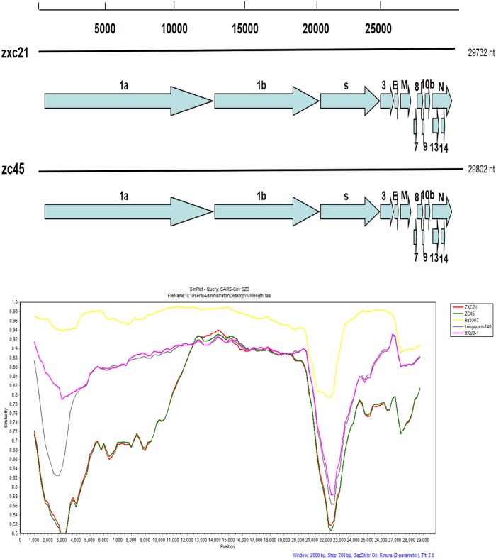
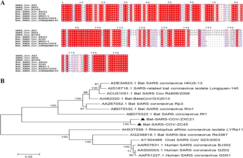
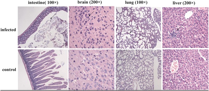
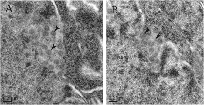
SARS coronavirus (SARS-CoV), the causative agent of the large SARS outbreak in 2003, originated in bats. Many SARS-like coronaviruses (SL-CoVs) have been detected in bats, particularly those that reside in China, Europe, and Africa. To further understand the evolutionary relationship between SARS-CoV and its reservoirs, 334 bats were collected from Zhoushan city, Zhejiang province, China, between 2015 and 2017. PCR amplification of the conserved coronaviral protein RdRp detected coronaviruses in 26.65% of bats belonging to this region, and this number was influenced by seasonal changes. Full genomic analyses of the two new SL-CoVs from Zhoushan (ZXC21 and ZC45) showed that their genomes were 29,732 nucleotides (nt) and 29,802 nt in length, respectively, with 13 open reading frames (ORFs). These results revealed 81% shared nucleotide identity with human/civet SARS CoVs, which was more distant than that observed previously for bat SL-CoVs in China. Importantly, using pathogenic tests, we found that the virus can reproduce and cause disease in suckling rats, and further studies showed that the virus-like particles can be observed in the brains of suckling rats by electron microscopy. Thus, this study increased our understanding of the genetic diversity of the SL-CoVs carried by bats and also provided a new perspective to study the possibility of cross-species transmission of SL-CoVs using suckling rats as an animal model.


Conflict of interest statement

All authors declare that they have no conflicts of interest.

Figures

Fig. 1
Fig. 1. A gene map of the two novel SL-CoVs and the recombination analysis of novel SL-CoVs with other SL- CoVs.
Similarity plots were conducted with SARS CoV SZ3 as the query and bat SL-CoVs, including Rs3367, Longquan-140, and HKU3-1, as potential parental sequences. The analysis was performed using the Kimura model, with a window size of 2000 base pairs and a step size of 200 base pairs
Fig. 2
Fig. 2. Characterization of S1 domains of the SARS CoV and SL-CoVs.
a Amino acid sequence comparison of the S1 subunit. The receptor-binding domain (aa 318–510) of SARS-CoV. b A phylogenetic analysis of the entire S1 amino acid sequences based on the neighbor-joining method. The SARS-CoV-GD01, BJ302, and GZ02 strains were isolated from patients of the SARS outbreak in 2003. The SARS-CoV SZ3 was identified from civets in 2003. Other bat-SL-CoVs were identified from bats in China.The sequences of SL-CoVs in this study are marked as filled triangles
Fig. 3
Fig. 3. Light microscopy observations of rat tissues infected with bat-SL-CoVs:
Sectioned brain, intestine, lung and liver tissues were sampled from rats infected with bat-SL-CoV ZC45
Fig. 4
Fig. 4. Transmission electron micrographs of infected rat brain tissues.
a, b CoV-like particles are considered SL-CoVs ZC45 in different locations of the infected rat brain tissues
Fig. 5
Fig. 5. Prediction of the antigenicity of the bat SL-CoV N protein.
a The predicted antigenicity for the N protein. b Amino acid sequence of the N protein. The high antigenicity portion is indicated in the red circle. The two synthesized polypeptides are indicated in red
Fig. 6
Fig. 6. Detection of N protein expression in infected rat tissues by western blotting.
Proteins from the following tissues were analyzed: rat brain from the control specimen (lane 1), intestinal tissue from bat ZC45 (lane 2), intestinal tissue from the infected rat (lane 3,6), lung tissue from the infected rat (lane 4,7), and brain tissue from the infected rat (lane 5,8)

References

    1. Cui J, et al. Evolutionary relationships between bat coronaviruses and their hosts. Emerg. Infect. Dis. 2007;13:1526–1532. doi: 10.3201/eid1310.070448. - DOI - PMC - PubMed
    1. Perlman S, Netland J. Coronaviruses post-SARS: update on replication and pathogenesis. Nat. Rev. Microbiol. 2009;7:439–450. doi: 10.1038/nrmicro2147. - DOI - PMC - PubMed
    1. Chinese SMEC. Molecular evolution of the SARS coronavirus during the course of the SARS epidemic in China. Science. 2004;303:1666–1669. doi: 10.1126/science.1092002. - DOI - PubMed
    1. Zhong NS, et al. Epidemiology and cause of severe acute respiratory syndrome (SARS) in Guangdong, People’s Republic of China, in February, 2003. Lancet. 2003;362:1353–1358. doi: 10.1016/S0140-6736(03)14630-2. - DOI - PMC - PubMed
    1. Marra MA, et al. The genome sequence of the SARS-associated coronavirus. Science. 2003;300:1399–1404. doi: 10.1126/science.1085953. - DOI - PubMed