. 2018 Sep 12;8(1):13704.
doi: 10.1038/s41598-018-32007-y.

IL-27 amplifies cytokine responses to Gram-negative bacterial products and Salmonella typhimurium infection

Affiliations

IL-27 amplifies cytokine responses to Gram-negative bacterial products and Salmonella typhimurium infection

C Petes et al. Sci Rep. .

Abstract

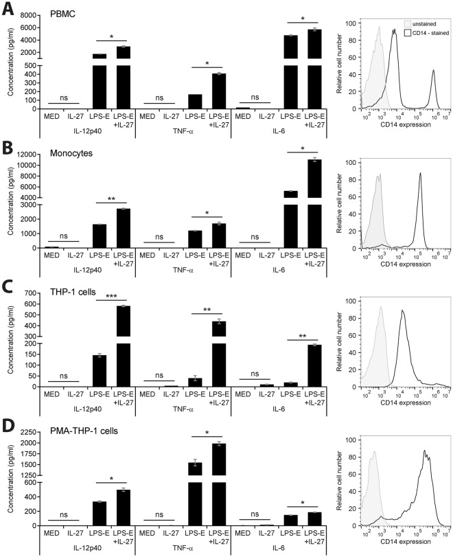
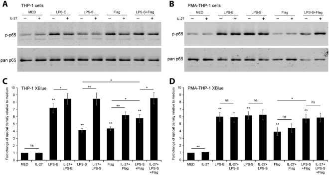
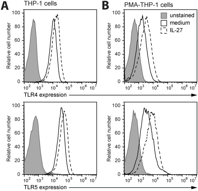
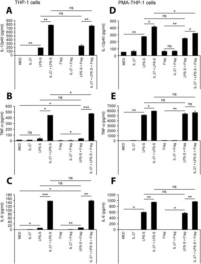
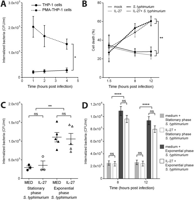
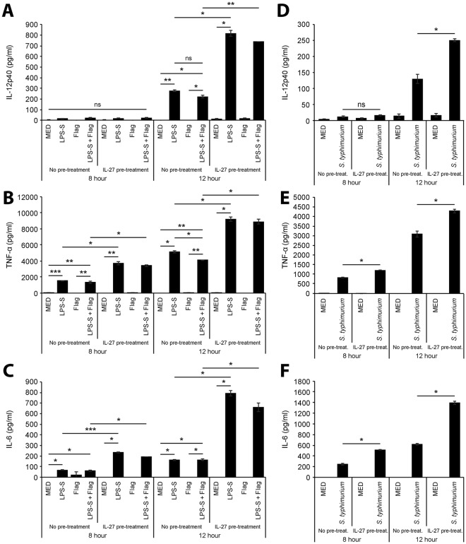
Cytokine responses from monocytes and macrophages exposed to bacteria are of particular importance in innate immunity. Focusing on the impact of the immunoregulatory cytokine interleukin (IL)-27 on control of innate immune system responses, we examined human immune responses to bacterial products and bacterial infection by E. coli and S. typhimurium. Since the effect of IL-27 treatment in human myeloid cells infected with bacteria is understudied, we treated human monocytes and macrophages with IL-27 and either LPS, flagellin, or bacteria, to investigate the effect on inflammatory signaling and cytokine responses. We determined that simultaneous stimulation with IL-27 and LPS derived from E. coli or S. typhimurium resulted in enhanced IL-12p40, TNF-α, and IL-6 expression compared to that by LPS alone. To elucidate if IL-27 manipulated the cellular response to infection with bacteria, we infected IL-27 treated human macrophages with S. typhimurium. While IL-27 did not affect susceptibility to S. typhimurium infection or S. typhimurium-induced cell death, IL-27 significantly enhanced proinflammatory cytokine production in infected cells. Taken together, we highlight a role for IL-27 in modulating innate immune responses to bacterial infection.


Conflict of interest statement

The authors declare no competing interests.

Figures

Figure 1
Figure 1
Co-stimulation with IL-27 enhanced LPS-induced cytokine production in human myeloid cells. Human PBMC (A), primary human monocytes (B), THP-1 cells (C), and PMA-THP-1 cells (D) were stimulated with LPS-E (100 ng/ml), IL-27 (50 ng/ml), or both LPS-E plus IL-27 concomitantly for 24 hours. IL-12p40, TNF-α and IL-6 levels (left panels) were measured in cell-free supernatants by ELISA. CD14 expression was measured by flow cytometry in resting cells (right panels). Histograms include CD14 expression and autofluorescence of each cell type (A–D, right panels). Data are representative of at least three different blood donors or independent replicate experiments showing mean and standard deviation of ELISA technical replicates from one experiment. Mann Whitney U tests were used for statistical analyses between pairs as indicated. ns = not significant; *p≤0.05; **p≤0.01; ***p≤0.001.
Figure 2
Figure 2
IL-27 altered TLR-mediated NF-κB/AP-1 activity in THP-1 monocytes, but not in PMA-THP-1 macrophages. THP-1 cells (A) and PMA-THP-1 cells (B) were stimulated with LPS-E (100 ng/ml), LPS-S (100 ng/ml), flagellin (Flag) (500 ng/ml), IL-27 (50 ng/ml), or combinations of TLR agonists plus IL-27 concomitantly as indicated for 15 minutes. Phosphorylation of NF-κBp65 subunit (p-p65) was presented using immunoblotting on whole cell lysates. Membranes were stripped and re-probed for pan NF-κBp65 (pan p65) as a loading control. Images shown are representative of three independent experiments. THP-1 XBlue cells (C) and PMA-THP-1 XBlue cells (D) were stimulated with LPS-E (100 ng/ml), LPS-S (100 ng/ml), flagellin (Flag) (500 ng/ml), IL-27 (50 ng/ml), or combinations of TLR agonists plus IL-27 concomitantly as indicated for 24 hours to allow for NF-κB/AP-1-induced SEAP production and secretion. SEAP production was quantified using a colorimetric QUANTI-Blue™ assay. Fold changes relative to medium controls (MED) were calculated. Data represent the mean and standard deviation of triplicate experiments. Mann Whitney U tests were used for statistical analyses between TLR agonists compared to MED (no lines) or between pairs as indicated (lines). ns = not significant; *p≤0.05; **p≤0.01.
Figure 3
Figure 3
Stimulation with IL-27 increased TLR4 and TLR5 expression in monocytes and macrophages. THP-1 cells (A) and PMA-THP-1 cells (B) were stimulated with or without IL-27 (50 ng/ml) for 16 hours. Cells were stained with anti-human TLR4 (top panels) or TLR5 (bottom panels) antibodies for receptor expression quantification by flow cytometry. Unstained cells were acquired to quantify autofluorescence of each cell type. Data are representative of at least three independent replicate experiments.
Figure 4
Figure 4
Simultaneous treatment with IL-27 results in elevated cytokine production in response to TLR4/TLR5 agonists. THP-1 cells (A–C) and PMA-THP-1 cells (D–F) were stimulated with LPS-S (100 ng/ml), flagellin (Flag) (500 ng/ml), IL-27 (50 ng/ml), or combinations of TLR agonists plus IL-27 concomitantly as indicated for 24 hours. IL-12p40 (top panels), TNF-α (middle panels), and IL-6 (bottom panels) levels were measured in cell-free supernatants by ELISA. Data are representative of at least three independent replicate experiments showing mean and standard deviation of ELISA technical replicates from one experiment. Mann Whitney U tests were used for statistical analyses between pairs as indicated. ns = not significant; *p≤0.05; **p≤0.01; ***p≤0.001.
Figure 5
Figure 5
IL-27 pre-treatment does not affect S. typhimurium internalization or infection-induced cell death. (A) THP-1 cells and PMA-THP-1 cells were infected with S. typhimurium (MOI: 10) for 1, 2, and 4 hours. Internalized bacteria were quantified (colony forming units (CFU)/ml) using the gentamicin protection assay. (B) PMA-THP-1 cells were treated with or without IL-27 (50 ng/ml) for 16 hours followed by infection with stationary phase S. typhimurium for 1.5, 8, and 12 hours. Propidium iodide staining was used to quantify cell death of PMA-THP-1 cells by flow cytometry. (C) PMA-THP-1 cells were treated with or without IL-27 (50 ng/ml) for 16 hours followed by infection with stationary phase or exponential phase S. typhimurium for 60 or 30 min, respectively. Gentamicin protection assay was used to determine the number of internalized bacteria (CFU/ml) immediately following infection. (D) PMA-THP-1 cells were treated with or without IL-27 (50 ng/ml) for 16 hours, then infected with stationary phase or exponential phase S. typhimurium for 60 or 30 min, respectively, followed by 8 and 12 hours of incubation. PMA-THP-1 associated bacteria were quantified (CFU/ml). Mann Whitney U tests were used for statistical analyses between individual pairs as indicated. One-way ANOVA tests were used for statistical analyses for comparisons between phases of S. typhimurium infection in cells treated with or without IL-27 as indicated. ns = not significant; *p≤0.05; **p≤0.01; ***p≤0.001; ****p≤0.0001.
Figure 6
Figure 6
Pre-treatment with IL-27 results in elevated cytokine production in response to S. typhimurium components and infection. PMA-THP-1 cells were treated with or without IL-27 (50 ng/ml) for 16 hours followed by LPS-S (100 ng/ml), flagellin (Flag) (500 ng/ml), or combinations of TLR agonists as indicated (AC) or stationary phase S. typhimurium (MOI: 10) (DF) for 8 or 12 hours. IL-12p40 (top panels), TNF-α (middle panels), and IL-6 (bottom panels) levels were measured in cell-free supernatants by ELISA. Data are representative of at least three independent replicate experiments showing mean and standard deviation of ELISA technical replicates from one experiment. Mann Whitney U tests were used for statistical analyses between pairs as indicated. ns = not significant; *p≤0.05; **p≤0.01; ***p≤0.001.

References

    1. Trinchieri G, Pflanz S, Kastelein RA. The IL-12 family of heterodimeric cytokines: new players in the regulation of T cell responses. Immunity. 2003;19:641–644. doi: 10.1016/S1074-7613(03)00296-6. - DOI - PubMed
    1. Pflanz S, et al. IL-27, a Heterodimeric Cytokine Composed of EBI3 and p28 Protein, Induces Proliferation of Naive CD4+ T Cells. Immunity. 2002;16:779–790. doi: 10.1016/S1074-7613(02)00324-2. - DOI - PubMed
    1. Villarino AV, Hunter CA. Biology of recently discovered cytokines: discerning the pro-and anti-inflammatory properties of interleukin-27. Arthritis Research And Therapy. 2004;6:225–225. doi: 10.1186/ar1227. - DOI - PMC - PubMed
    1. Pflanz S, et al. WSX-1 and glycoprotein 130 constitute a signal-transducing receptor for IL-27. The Journal of Immunology. 2004;172:2225–2231. doi: 10.4049/jimmunol.172.4.2225. - DOI - PubMed
    1. Taga T, Kishimoto T. Gp130 and the interleukin-6 family of cytokines. Annu Rev Immunol. 1997;15:797–819. doi: 10.1146/annurev.immunol.15.1.797. - DOI - PubMed

Publication types