

Patterns of posterior ocular complications in myopic eyes of Indian population
- PMID: 30209314
- PMCID: PMC6135820
- DOI: 10.1038/s41598-018-29536-x
Patterns of posterior ocular complications in myopic eyes of Indian population
Abstract
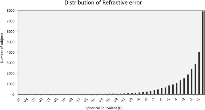
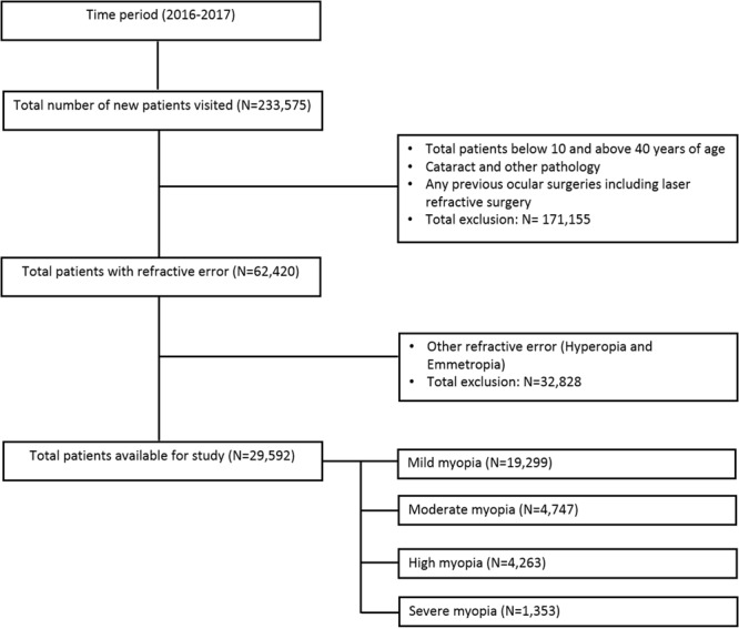
This is a retrospective study aimed to investigate the patterns of myopic fundus complications in Indian children and young adults. Electronic medical records of 29,592 patients, aged 10-40 years, who visited L V Prasad Eye Institute between 1st January to 31st December 2016 were analysed in the study. Data such as age, gender, refractive error and various pathologic lesions of posterior globe were considered for analysis. Among all the patients with different types of refractive errors, myopia was found in 47.4%, high myopia in 6.8% and pathologic myopia in 2.2%. There was no trend of the increased prevalence of pathologic myopia with increasing age, except for a significant difference between the children aged 10-15 years (2.7%) and those aged more than 15 years (>4%). . Although, the overall pattern of pathologic lesions was similar across different grades of myopia (2.5% in low myopes vs. 2.2% in severe myopes), lesions like staphyloma and retinal detachment increased with increasing degree of myopia. The proportion of pathologic lesions across different grades of myopia suggests the necessity for careful peripheral fundus examinations irrespective of the degree of myopia for better management and prognostic purposes.
Conflict of interest statement
The authors declare no competing interests.
Figures
References
MeSH terms
LinkOut - more resources
Full Text Sources
Other Literature Sources
Medical
