. 2019 Feb;92(1094):20180157.
doi: 10.1259/bjr.20180157. Epub 2018 Dec 17.

Combined application of isotropic three-dimensional fast spin echo (3D-FSE-Cube) with 2-point Dixon fat/water separation (FLEX) and 3D-FSE-cube in MR dacryocystography

Affiliations

Combined application of isotropic three-dimensional fast spin echo (3D-FSE-Cube) with 2-point Dixon fat/water separation (FLEX) and 3D-FSE-cube in MR dacryocystography

Ping Liu et al. Br J Radiol. 2019 Feb.

Abstract

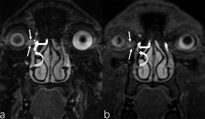
Objective:: To evaluate the image quality of magnetic resonance dacryocystography (MRD) using three-dimesional fast spin-echo -Cube (3D-FSE-Cube) and 3D-FSE-Cube-Flex sequences to examine the lacrimal drainage system (LDS).

Methods:: 21 healthy volunteers underwent 3D-FSE-Cube and 3D-FSE-Cube-Flex MRD after topical administration of compound sodium chloride eye drops. Two radiologists assessed LDS images in a blinded fashion. The signal-to-noise ratio of fluid-filling and the contrast-to-noise ratio of fluid-turbinate were compared between the two sequences. Overall image quality, sharpness, artefacts, visualization of anatomical structures, and visibility of LDS segments were also compared.

Results:: Overall image quality, visualization of anatomic structures, and artefact were significantly better on 3D-FSE-Cube-Flex MRD (p < 0.001, respectively). when compared to 3D-FSE-Cube. 3D-FSE-Cube showed lower fluid-filling signal-to-noise ratio and fluid-inferior turbinate CNR (all p < 0.001). In comparison with 3D-FSE-Cube-Flex, 3D-FSE-Cube produced superior visibility of the upper drainage segments (superior canaliculi, p = 0.003; common canaliculus, p = 0.033; inferior canaliculi, p < 0.001), but inferior in lower-LDS visibility (lacrimal sac, p = 0.001; nasolacrimal duct, p < 0.001). There was no difference in the total number of segments visualized per LDS between the two sequences (p = 0.068).

Conclusions:: 3D-FSE-Cube-Flex demonstrated superior image quality and visibility of the lower LDS segments. 3D-FSE-Cube showed an advantage in visualizing the upper LDS segments. The combination of these sequences can improve LDS visibility.

Advances in knowledge:: 3D-FSE-Cube-Flex provides robust water & fat separation and mitigates lower LDS-associated inhomogeneity artefacts. 3D-FSE-Cube shows optimal upper LDS visualization. The combined application of these sequences is a non-invasive and effective method for assessing LDS disease.


Conflict of interest statement

Competing interests: The authors Ping Liu and Qiuxia Wang contributed equally to the work.

Figures

Figure 1.
Figure 1.
Examples of ROI placement for filling-fluid and noise. ROI, region of interest.
Figure 2.
Figure 2.
3D-FSE-Cube-Flex for a subject in the (a) oblique coronal, (b) and (c) bilateral oblique sagittal planes. Example of 3D-FSE-Cube for a subject reformatted in the (d) oblique coronal, (e) and (f) bilateral oblique sagittal planes. The attached liquid delineates the profile of the bilateral LS and NLD, with the latter sequence showing a sharper boundary. 3D-FSE, three-dimensional-fast spin-echo; LS, lacrimal sac; NLD, nasolacrimal duct.
Figure 3.
Figure 3.
An example of a subject with (A) reformatted oblique coronal 3D-FSE-Cube MRD that clearly delineates the SC and IC (straight arrows), CC (slender white arrow), LS (thick bent arrow), and NLD (curved arrow); and, (B) reformatted oblique coronal 3D-FSE-Cube-Flex MRD, with the CC not visible, as well as the SC and IC are faintly displayed (straight arrows).
Figure 4.
Figure 4.
The bar chart at the top shows the comparison of SNR of fluid-filling and fluid-inferior turbinate CNR between 3D-FSE-Cube MRD and 3D-FSE-Cube-Flex MRD (mean ± SD). There is a trend toward higher fluid SNR with the 3D-FSE-Cube-Flex over 3D-FSE-Cube, but a contrary tendency in that of CNR in terms of the MRD (p < 0.001, respectively). The table below the histogram displays quantitative assessment. Data was presented as mean ± SD and based on all images were obtained with fat-suppression (On 3D-FSE-Cube-Flex, it is mainly the water-only image). p < 0.05 was considered statistically significant. 3D-FSE, three-dimensional-fast spin-echo; CNR, contrats-to-noise ratio; MRD, magnetic resonance dacryocystography; NLD, nasolacrimal duct; SD, standard deviation; SNR, signal-to-noise ratio.
Figure 5.
Figure 5.
The histogram displays comparison of the segment visualization scores between 3D-FSE-Cube MRD and 3D-FSE-Cube-Flex MRD, with a trend toward higher scores with superior LDSs for 3D-FSE-Cube MRD, with SC (p = 0.003), IC (p < 0.001), and CC (p = 0.033) being significant. Lower NLDs and the total sections per subject showed a trend with lower rating scores. Data in the table is presented as Mean ± SD and based on all images were obtained with fat-suppression (the water-only images from 3D-FSE-Cube-Flex MRD). The measure of visualization for each ductal section range form 0–3: Grade 0, invisible and no fluid filling; Grade 1, less than half of the passage can be discerned and filled; Grade 2, more than half of the passage was filled and can be faintly visualized; Grade 3, clear and full visualization. p < 0.05 was considered statistically significant. 3D-FSE, three-dimensional-fast spin-echo; CC, common canaliculus; IC, inferior canaliculus; LS, lacrimal sac; MRD, magnetic resonance dacryocystography; NLD, nasolacrimal duct; SC, superior canaliculus; SD, standard deviation.
Figure 6.
Figure 6.
Example image of a 22-year-old female volunteer, in which the maxillary sinus was filled with gas. The reformatted coronal (a) and oblique sagittal planes (c) of NLD (swallowtail arrows) on 3D-FSE-Cube-Flex; and reformatted coronal (b) and oblique sagittal planes (d) of NLD on 3D-FSE-Cube for the same subject. 3D-FSE-Cube-Flex MRD demonstrated minimal and serrated artefacts, but there were more columnar artefacts with 3D-FSE-Cube. 3D-FSE, three-dimensional-fast spin-echo; NLD, nasolacrimal duct.

References

    1. Busse H, Müller KM, Kroll P, Radiological KP, Findings.pdf H. Radiological and histological findings of the lacrimal passages of newborns. Arch Ophthalmol 1980; 98: 528–32. doi: 10.1001/archopht.1980.01020030524019 - DOI - PubMed
    1. Maliborski A, Różycki R. Diagnostic imaging of the nasolacrimal drainage system. Part I. Radiological anatomy of lacrimal pathways. Physiology of tear secretion and tear outflow. Med Sci Monit 2014; 20: 628–38. doi: 10.12659/MSM.890098 - DOI - PMC - PubMed
    1. Caldemeyer KS, Stockberger SM, Broderick LS. Topical contrast-enhanced CT and MR dacryocystography: imaging the lacrimal drainage apparatus of healthy volunteers. AJR Am J Roentgenol 1998; 171: 1501–4. doi: 10.2214/ajr.171.6.9843278 - DOI - PubMed
    1. Yoshikawa T, Hirota S, Sugimura K. Topical contrast-enhanced magnetic resonance dacryocystography. Radiat Med 2000; 18: 355–62. - PubMed
    1. Somma F, d’Agostino V, Tortora F, Serra N, Sorrentino G, Piscitelli V, et al. . Magnetic resonance imaging in the pre-operative evaluation of obstructive epiphora: true-FISP and VIBE vs gadolinium. Radiol Med 2017; 122: 123–30. doi: 10.1007/s11547-016-0696-4 - DOI - PubMed

MeSH terms

Substances