

Aortic Mural Thrombus in the Non-atherosclerotic Aorta of Patients with Multiple Hypercoagulable Factors
- PMID: 30210102
- PMCID: PMC6395124
- DOI: 10.2169/internalmedicine.0691-17
Aortic Mural Thrombus in the Non-atherosclerotic Aorta of Patients with Multiple Hypercoagulable Factors
Abstract
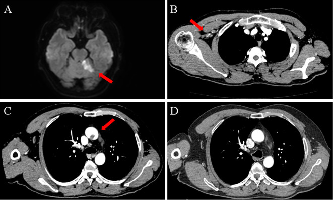
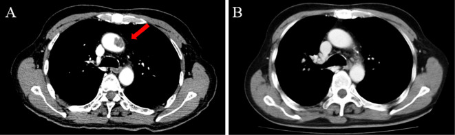
An aortic mural thrombus (AMT) on a non-atherosclerotic wall is a rare but important cause of arterial thromboembolism. We herein report two cases of AMT in the thoracic aorta. Both showed multiple hypercoagulable factors (case 1: protein S deficiency and positive finding of anti-cardiolipin antibody; case 2: protein C deficiency, gastric cancer, and cisplatin-based chemotherapy) and were successfully treated with anticoagulation. Hypercoagulable states, including malignancy, can influence the formation of AMT; therefore, the accurate assessment of a hypercoagulable condition is necessary when we encounter patients with AMT.
Keywords: anticoagulant therapy; aortic thrombus; arterial embolism; hypercoagulable disorder.
Conflict of interest statement
Figures
References
-
- Abbott WM, Maloney RD, McCabe CC, Lee CE, Wirthlin LS. Arterial embolism: a 44 year perspective. Am J Surg 143: 460-464, 1982. - PubMed
-
- Machleder HI, Takiff H, Lois JF, Holburt E. Aortic mural thrombus: An occult source of arterial thromboembolism. J Vasc Surg 4: 473-478, 1986. - PubMed
-
- Tunick PA, Kronzon I. Atheromas of the thoracic aorta: clinical therapeutic update. J Am Coll Cardiol 35: 545-554, 2000. - PubMed
-
- Cassella CR, Jagoda A. Atherosclerotic disease of the aortic arch as a risk factor for recurrent ischemic stroke. N Engl J Med 334: 1216-1221, 1996. - PubMed
-
- Laperche T, Laurian C, Roudaut R, Steg PG. Mobile thromboses of the aortic arch without aortic debris. Circulation 96: 288-294, 1997. - PubMed
Publication types
MeSH terms
Substances
LinkOut - more resources
Full Text Sources
Other Literature Sources
Medical