. 2018 Aug 24:11:5171-5181.
doi: 10.2147/OTT.S160066. eCollection 2018.

Prognostic value of SOX2, Cyclin D1, P53, and ki-67 in patients with esophageal squamous cell carcinoma

Affiliations

Prognostic value of SOX2, Cyclin D1, P53, and ki-67 in patients with esophageal squamous cell carcinoma

Hui Wang et al. Onco Targets Ther. .

Abstract

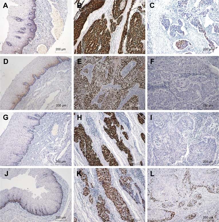
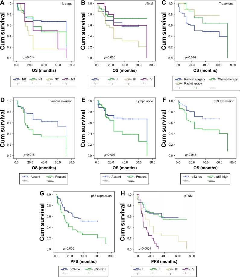
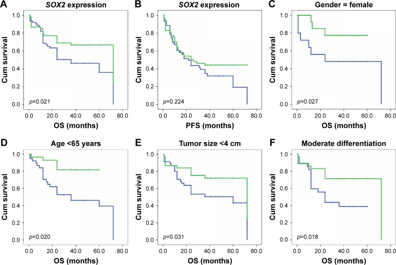
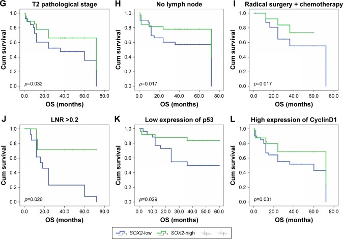
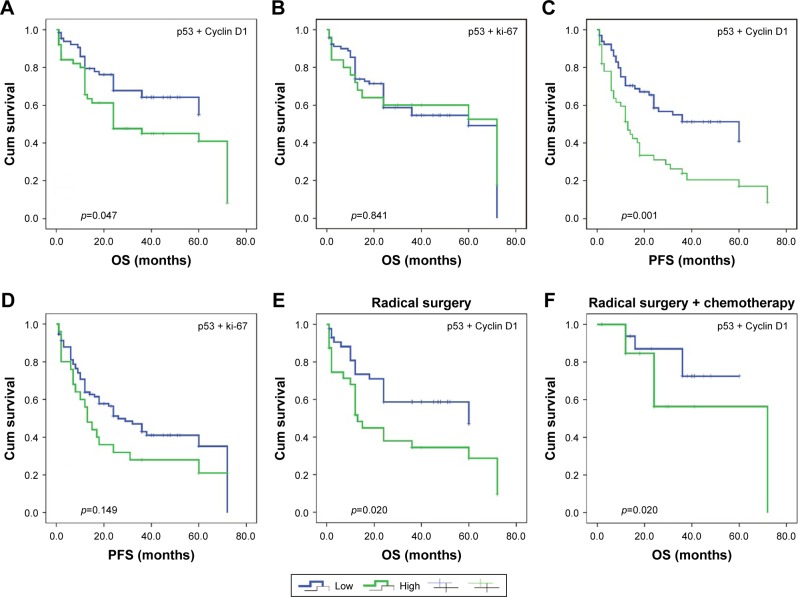
In this study, we evaluated SOX2, Cyclin D1, p53, and ki-67 expression immunohistochemically in 117 samples of surgically resected esophageal squamous cell carcinoma (ESCC) and matched normal tumor adjacent tissues and correlated the expression with clinicopathological finding and patient survival. Lymph node metastasis was observed in 36.8% of patients, and organ metastasis was observed in 17.9%. We detected high expression of SOX2, Cyclin D1, p53, and ki-67 in 46.1%, 70.1%, 54.7%, and 32.5% of ESCC tissues, respectively. SOX2 is localized in the tumor cell nuclei, and its expression was significantly associated with N stage (p=0.034) and differentiation (p=0.003) and ki-67 expression (p=0.001), whereas increased Cyclin D1 expression was correlated with high p53 (p=0.015). With regard to survival, we found that ESCC patients with high SOX2 expression had significantly better survival time than those with low SOX2 expression (p=0.021). A multivariate Cox analysis revealed that therapy and high p53 expression and venous invasion were independent predictors of unfavorable prognosis in overall survival (p=0.039, p=0.004, and p=0.023, respectively). Furthermore, higher T stage, clinical stage (pTNM), venous invasion, and high p53 expression were independent predictors of a worse progression-free survival. Notably, co-overexpression of p53 and Cyclin D1 was significantly correlated with poor overall survival and progression-free survival (p=0.029 and p=0.0227, respectively). Therefore, SOX2 might be considered as a potential prognostic indicator and a potential target for therapeutic targets in ESCC. p53 staining and combined p53 and Cyclin D1 expression had significantly unfavorable prognostic value for patients with ESCC. These findings provide more insight into ESCC; thus, further investigations into molecular mechanisms of drug resistance are essential.

Keywords: Cyclin D1; SOX2; esophageal squamous cell carcinoma; ki-67; p53; prognosis.


Conflict of interest statement

Disclosure The authors report no conflicts of interest in this work.

Figures

Figure 1
Figure 1
IHC staining of SOX2, Cyclin D1, P53, and KI-67 in ESCC tissues. Notes: (A) In normal esophagus mucosa, SOX2 is localized to the basal layer (100×). (B) Strong positive expression of SOX2 in ESCC (100×). (C) SOX2 was expressed at a low level in ESCC (100×). (D) Cyclin D1 expression in normal esophagus epithelium (100×). (E) High expression of Cyclin D1 in ESCC (100×). (F) Low expression of Cyclin D1 in ESCC (100×). (G) P53 was expressed in adjacent normal esophagus mucosa (100×). (H) P53 was strongly positive in ESCC (100×). (I) P53 was negative in ESCC (100×). (J) In normal esophagus mucosa, ki-67 is localized to the basal layer (100×). (K) Ki-67 upregulation in ESCC (100×). (L) Low expression of ki-67 in ESCC (100×). Abbreviations: ESCC, esophageal squamous cell carcinoma; IHC, immunohistochemistry.
Figure 2
Figure 2
Blank staining of SOX2, Cyclin D1, p53, and Ki-67 in ESCC tissues (100×). Notes: (A) Negative control of SOX2 in esophagus mucosa; (B) Negative control of SOX2 in ESCC; (C) Negative control of Cyclin D1 in esophagus mucosa; (D) Negative control of Cyclin D1 in ESCC; (E) Negative control of p53 in esophagus mucosa; (F) Negative control of p53 in ESCC; (G) Negative control of Ki-67 in esophagus mucosa; and (H) Negative control of Ki-67 in ESCC. Abbreviation: ESCC, esophageal squamous cell carcinoma.
Figure 3
Figure 3
OS and PFS analysis of patients with ESCC using the Kaplan–Meier method. Notes: OS according to (A) N stage; (B) pTNM; (C) treatment; (D) venous invasion; (E) lymph node; (F) p53 expression (low vs high). PFS according to (G) p53 expression (low vs high); (H) pTNM. Abbreviations: OS, overall survival; PFS, progression-free survival; ESCC, esophageal squamous cell carcinoma.
Figure 4
Figure 4
OS and PFS based on SOX2 expression. Notes: (A) OS; (B) PFS. The OS of ESCC patients was analyzed using Kaplan–Meier and stratified by SOX2 expression level. (C) Female; (D) age <65 years; (E) tumor size <4 cm; (F) moderate differentiation; (G) pathological stage: T2; (H) absent lymph node metastasis; (I) receiving both radical surgery and chemotherapy; (J) LNRs ≥0.2; (K) low expression of p53; and (L) high expression of Cyclin D1. Abbreviations: LNR, lymph node ratio; OS, overall survival; PFS, progression-free survival; ESCC, esophageal squamous cell carcinoma.
Figure 4
Figure 4
OS and PFS based on SOX2 expression. Notes: (A) OS; (B) PFS. The OS of ESCC patients was analyzed using Kaplan–Meier and stratified by SOX2 expression level. (C) Female; (D) age <65 years; (E) tumor size <4 cm; (F) moderate differentiation; (G) pathological stage: T2; (H) absent lymph node metastasis; (I) receiving both radical surgery and chemotherapy; (J) LNRs ≥0.2; (K) low expression of p53; and (L) high expression of Cyclin D1. Abbreviations: LNR, lymph node ratio; OS, overall survival; PFS, progression-free survival; ESCC, esophageal squamous cell carcinoma.
Figure 5
Figure 5
OS according to combined p53-Cyclin D1 expression. Notes: (A) Combined p53-Ki-67 expression; (B) PFS based on combined p53-Cyclin D1 expression; (C) coexpression of p53 and Ki-67; (D) OS and PFS based on coexpression of p53 and Cyclin D1 stratified by treatment; (E) receiving radical surgery treatment; and (F) receiving both radical surgery and chemotherapy. Abbreviations: OS, overall survival; PFS, progression-free survival.

Similar articles

Cited by

References

    1. Rustgi AK, El-Serag HB. Esophageal carcinoma. N Engl J Med. 2014;371(26):2499–2509. - PubMed
    1. Napier KJ, Scheerer M, Misra S. Esophageal cancer: a review of epidemiology, pathogenesis, staging workup and treatment modalities. World J Gastrointest Oncol. 2014;6(5):112–120. - PMC - PubMed
    1. Torre LA, Bray F, Siegel RL, Ferlay J, Lortet-Tieulent J, Jemal A. Global cancer statistics, 2012. CA Cancer J Clin. 2015;65(2):87–108. - PubMed
    1. Wang H, Deng F, Liu Q, Ma Y. Prognostic significance of lymph node metastasis in esophageal squamous cell carcinoma. Pathol Res Pract. 2017;213(7):842–847. - PubMed
    1. Pennathur A, Gibson MK, Jobe BA, Luketich JD. Oesophageal carcinoma. Lancet. 2013;381(9864):400–412. - PubMed