Review
. 2018 Aug 29:10:29.
doi: 10.3389/fnsyn.2018.00029. eCollection 2018.

Plasticity of Spine Structure: Local Signaling, Translation and Cytoskeletal Reorganization

Affiliations
Review

Plasticity of Spine Structure: Local Signaling, Translation and Cytoskeletal Reorganization

Yoshihisa Nakahata et al. Front Synaptic Neurosci. .

Abstract

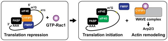
Dendritic spines are small protrusive structures on dendritic surfaces, and function as postsynaptic compartments for excitatory synapses. Plasticity of spine structure is associated with many forms of long-term neuronal plasticity, learning and memory. Inside these small dendritic compartments, biochemical states and protein-protein interactions are dynamically modulated by synaptic activity, leading to the regulation of protein synthesis and reorganization of cytoskeletal architecture. This in turn causes plasticity of structure and function of the spine. Technical advances in monitoring molecular behaviors in single dendritic spines have revealed that each signaling pathway is differently regulated across multiple spatiotemporal domains. The spatial pattern of signaling activity expands from a single spine to the nearby dendritic area, dendritic branch and the nucleus, regulating different cellular events at each spatial scale. Temporally, biochemical events are typically triggered by short Ca2+ pulses (~10-100 ms). However, these signals can then trigger activation of downstream protein cascades that can last from milliseconds to hours. Recent imaging studies provide many insights into the biochemical processes governing signaling events of molecular assemblies at different spatial localizations. Here, we highlight recent findings of signaling dynamics during synaptic plasticity and discuss their roles in long-term structural plasticity of dendritic spines.

Keywords: CaMKII; FRET FLIM; LTP; actin cytoskeleton; calcium signaling; dendritic spine; small GTPase; synaptic plasticity.


Figures

Figure 1
Figure 1
Schematic of actin and actin-binding proteins (ABPs) in a dendritic spine. Filamentous actin (F-actin) is formed by polymerization of globular actin (G-actin). The constant process of polymerization of ATP-bound G-actin (magenta oval) at barbed (plus) end and depolymerization of ADP-bound G-actin (cyan oval) at pointed (minus) end is called actin tread-milling. Profilin (yellow square) binds to monomeric G-actin and accelerates exchange of its nucleotide from ADP to ATP, as the result enhancing actin polymerization. ADF/cofilin (black oval) binds to ADP-bound actin and accelerates actin depolymerization at a low concentration. Arp2/3 complex (white oval) nucleates actin branching. The function of Arp2/3 complex is activated by Wiskott-Aldrich syndrome family protein (WASP) and inhibited by WASP family verprolin-homologous protein (WAVE). Epidermal growth factor receptor kinase substrate 8 (Eps8), binds to barbed-end and stabilizes actin filaments. Cross-linking proteins including actinin, CaMKIIβ and drebrin stabilize F-actin and form actin network. Active vesicular transport along F-actin is regulated by myosin.
Figure 2
Figure 2
Intracellular signal regulation during structural long-term potentiation (LTP). (A) Signaling pathways controlling actin binding proteins (ABPs) in dendritic spines. Black arrows represent downstream activation and red lines represent downstream inhibition. (B) Different contributions of small GTPase activations in transient and sustained structural LTP (sLTP) in stimulated spines. Red dot represents a single spine stimulation by glutamate uncaging. (C) Schematic time course of small GTPase activation profiles in stimulated spines (Murakoshi et al., ; Oliveira and Yasuda, ; Hedrick et al., 2016).
Figure 3
Figure 3
Intracellular and extracellular factors that regulate spine structural plasticity. Activity-dependent autocrine brain-derived neurotrophic factor (BDNF)-TrkB signaling activates Cdc42 and Rac1 in single spines. Ca2+ influx through NMDA receptors or voltage gated Ca2+ channels (VGCCs) activates CaMKIIα and its downstream signaling including Cdc42, Rac1, RhoA and Ras. Activation of Rac1 and Cdc42 are regulated by autocrine BDNF-TrkB signaling. Activities of RhoA, Rac1 and Ras spread to dendritic shaft and adjacent spines from stimulated spines. MMP-9 and TIMP-1 are also released from the postsynaptic cells. In addition, Ca2+ elevation induces lysosomal fusion with the plasma membrane, releasing Cathepsin B to outside of the cell. Extracellular Cathepsin B cleaves the tissue inhibitor of metalloproteinases-1 (TIMP-1), an endogenous inhibitor of the matrix metalloprotease 9 (MMP-9). Disinhibited MMP-9 cleaves cell adhesion molecules (CAMs) and the extracellular matrix (ECM), which facilitate structural remodeling of spines. Broken arrows represents signal spreading.
Figure 4
Figure 4
Schematic diagram of local translation in dendrites. mRNA is transported in the ribonucleoprotein (RNP) particle, which includes mRNA, translation initiation factors, ribosomal subunits, RNA binding proteins (RBPs) and microRNA (miRNA). Association with RNA binding proteins (RBPs) at 3’-UTR (representing as AAA) and eukaryotic translation initiation factor 4E (eIF4E) at 5’-cap (representing as m7G). RBP binding is critical for dendritic trafficking of mRNA, while repressing translation. Cap-dependent translation initiation is regulated by the interaction of mRNA with eIF4E binding protein (4E-BP). Kinesin and dynein actively transport RNP toward anterograde and retrograde directions along microtubules, respectively. Unloading of β-actin mRNA and translation initiation are simultaneously regulated at the base of stimulated spines. Newly synthesized β-actin translocates to the peripheral region of the stimulated spine head. Warm color depicts dynamic actin pools. Newly synthesized Arc preferentially increases in non-stimulated spines and changing cytoskeletal dynamics via interacting ABPs including WAVE1, ADF/cofilin, CaMKIIβ and drebrin A. Arc promotes AMPA receptor (AMPAR) endocytosis through interaction to dynamin-2 (Dyn2).
Figure 5
Figure 5
Dual roles ofcytoplasmic fragile X mental retardation protein (FMRP) interacting protein 1 (CYFIP1) for translation initiation and rapid actin remodeling. CYFIP1 makes FMRP-CYFIP1-eIF4E complex on mRNA and plays a role as a non-canonical eukaryotic initiation factor 4E (eIF4E) binding protein (4E-BP) which represses the translation initiation by interfering the association of eukaryotic initiation factor 4G (eIF4G). Binding of active form Rac1 (GTP-Rac1) dissociates CYFIP1 from the mRNA complex and initiates the translation. Dissociated CYFIP1 from mRNA complex forms WAVE complex, which promotes actin nucleation and branching via activating Arp2/3 complex. PABP, poly (A)-binding protein.

Similar articles

Cited by

References

    1. Ackermann M., Matus A. (2003). Activity-induced targeting of profilin and stabilization of dendritic spine morphology. Nat. Neurosci. 6, 1194–1200. 10.1038/nn1135 - DOI - PubMed
    1. Andrianantoandro E., Pollard T. D. (2006). Mechanism of actin filament turnover by severing and nucleation at different concentrations of ADF/cofilin. Mol. Cell 24, 13–23. 10.1016/j.molcel.2006.08.006 - DOI - PubMed
    1. Arber S., Barbayannis F. A., Hanser H., Schneider C., Stanyon C. A., Bernard O., et al. . (1998). Regulation of actin dynamics through phosphorylation of cofilin by LIM-kinase. Nature 393, 805–809. 10.1038/31729 - DOI - PubMed
    1. Ashley J., Cordy B., Lucia D., Fradkin L. G., Budnik V., Thomson T. (2018). Retrovirus-like Gag protein Arc1 binds RNA and traffics across synaptic boutons. Cell 172, 262.e11–274.e11. 10.1016/j.cell.2017.12.022 - DOI - PMC - PubMed
    1. Barria A., Muller D., Derkach V., Griffith L. C., Soderling T. R. (1997). Regulatory phosphorylation of AMPA-type glutamate receptors by CaM-KII during long-term potentiation. Science 276, 2042–2045. 10.1126/science.276.5321.2042 - DOI - PubMed

LinkOut - more resources