

eCollection 2017 Nov-Dec.
Microglia: The Brain's First Responders
- PMID: 30210663
- PMCID: PMC6132046
Item in Clipboard
Microglia: The Brain's First Responders
Cerebrum.
.
Abstract
New knowledge about microglia is so fresh that it's not even in the textbooks yet. Microglia are cells that help guide brain development and serve as its immune system helpers by gobbling up diseased or damaged cells and discarding cellular debris. Our authors believe that microglia might hold the key to understanding not just normal brain development, but also what causes Alzheimer's disease, Huntington's disease, autism, schizophrenia, and other intractable brain disorders.
Figures
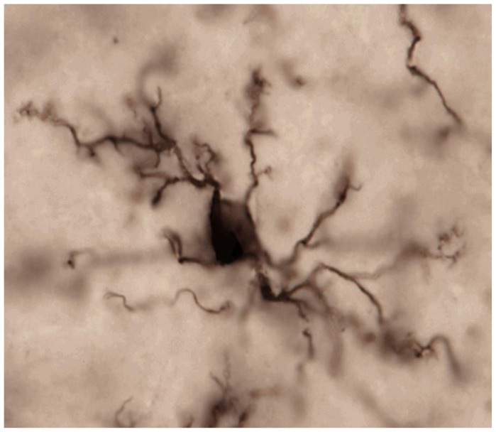
Microglia in the mature, healthy brain exhibit small cell bodies and multiple long, thin processes (arms) that they use to constantly scan and survey their local environments within brain tissue. Photo credit S. Bilbo.
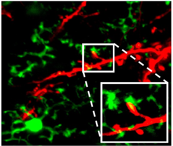
Microglia dynamically interact with synaptic elements in the healthy brain. Two-photon imaging in the olfactory bulb of adult mice shows processes of CX3CR1-GFP-positive microglia connecting to tdTomato-labeled neurons. Reprinted with permission from Jenelle Wallace at Harvard University (Hong and Stevens, 201620).
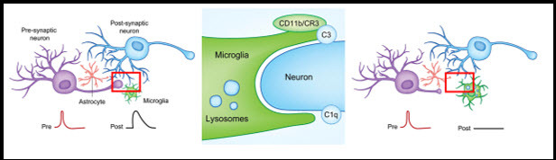
Synaptically coupled (i.e. communicating) neurons are under constant surveillance by glial cells, including microglia. If a neuronal synapse becomes “tagged” with complement protein C3, microglia recognize the tag with their C3 receptor (CR3/CD11b). This signal tells the microglia to engulf, or phagocytosis, and degrade the synapse. After microglial synaptic pruning, the eliminated synapse changes the way neurons communicate. Adapted from Lacagnina et al., 2017.
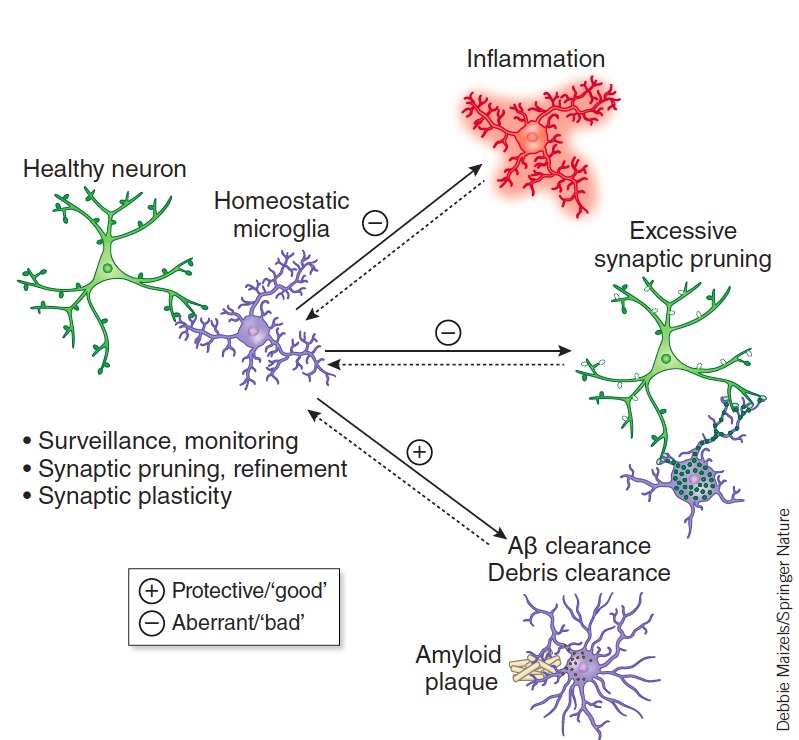
Microglia have complex roles that are both beneficial and detrimental to disease pathogenesis including engulfing or degrading toxic proteins (i.e., amyloid plaques) and promoting neurotoxicity through excessive inflammatory cytokine release. Aberrations in microglia’s normal homeostatic functions (Surveillance, synaptic pruning and plasticity) may also contribute to excessive synapse loss and cognitive dysfunction in AD and other diseases. Salter and Stevens 2016 with permission.
References
-
- Deverman BE, Patterson PH. Cytokines and CNS development. Neuron. 2009;64(1):61–78. - PubMed
-
- Sierra A, de Castro F, Del Rio-Hortega J, Rafael Iglesias-Rozas J, Garrosa M, Kettenmann H. The “Big-Bang” for modern glial biology: Translation and comments on Pio del Rio-Hortega 1919 series of papers on microglia. Glia. 2016;64(11):1801–1840. - PubMed
LinkOut - more resources
Full Text Sources