. 2018 Sep 11:6:28.
doi: 10.1038/s41413-018-0028-5. eCollection 2018.

Mandible exosomal ssc-mir-133b regulates tooth development in miniature swine via endogenous apoptosis

Affiliations

Mandible exosomal ssc-mir-133b regulates tooth development in miniature swine via endogenous apoptosis

Ye Li et al. Bone Res. .

Abstract

Signal transduction between different organs is crucial in the normal development of the human body. As an important medium for signal communication, exosomes can transfer important information, such as microRNAs (miRNAs), from donors to receptors. MiRNAs are known to fine-tune a variety of biological processes, including maxillofacial development; however, the underlying mechanism remains largely unknown. In the present study, transient apoptosis was found to be due to the expression of a miniature swine maxillofacial-specific miRNA, ssc-mir-133b. Upregulation of ssc-mir-133b resulted in robust apoptosis in primary dental mesenchymal cells in the maxillofacial region. Cell leukemia myeloid 1 (Mcl-1) was verified as the functional target, which triggered further downstream activation of endogenous mitochondria-related apoptotic processes during tooth development. More importantly, mandible exosomes were responsible for the initial apoptosis signal. An animal study demonstrated that ectopic expression of ssc-mir-133b resulted in failed tooth formation after 12 weeks of subcutaneous transplantation in nude mice. The tooth germ developed abnormally without the indispensable exosomal signals from the mandible.


Conflict of interest statement

The authors declare no competing interests.

Figures
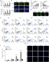
Fig. 1
Fig. 1
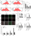
Ssc-mir-133b was highly related to cell apoptosis in the early stages of premolar development. a Relative expression of ssc-mir-133b in premolar dental epithelium and mesenchyme using qPCR. U6 was used as a control. Average ssc-mir-133b expression levels in premolar the dental mesenchyme were significantly higher than in the dental epithelium (upper panel). Similarly, the ssc-mir-133b expression levels in primary premolar dental mesenchyme cells were significantly higher than in dental epithelium cells (lower panel). ***P < 0.001. b Apoptosis was measured with a TUNEL assay in E40, E50, and E60 premolars. Scale bar = 200 μm. Enamel knots (EK, red dotted circle). c Relative ssc-mir-133b expression levels at 48 h after transfection of different MOIs of ssc-mir-133b lentiviral vectors and the efficiency of ssc-mir-133b lentiviral vector transfection. Average ssc-mir-133b expression levels after transfection with ssc-mir-133b overexpression lentiviral vector (upper left). average ssc-mir-133b expression levels after transfection with ssc-mir-133b inhibition lentiviral vector (upper right). ***P < 0.001. The efficiency of the ssc-mir-133b lentiviral vector (lower). d Cells dually stained with annexin-FITC/PI were investigated by flow cytometry analysis. Q1 quadrant represents the living cells; Q2 quadrant represents the early apoptotic cells; Q3 quadrant represents the late apoptotic cells; and Q4 quadrant represents the dead cells. e Statistical analysis of flow cytometry results. ***P < 0.001 and **P < 0.01. f Transfected premolar mesenchymal cells were stained with Hoechst 33342 on coverslips. A fluorescence microscope was used to photograph the results. Bar = 50 μm
Fig. 2
Fig. 2
Mcl-1 is the downstream target gene of ssc-mir-133b during early premolar development. a In situ hybridization with DIG-labeled Mcl-1 cDNA in E40, E50, and E60 premolars. Scale bar = 200 μm. Enamel knots (EK, red dotted circle). b The Mcl-1 3′-UTR harbors one ssc-mir-133b-specific binding site. c Relative ssc-mir-133b expression levels at 48 h after transfection of ssc-mir-133b mimics and inhibitors. Relative ssc-mir-133b levels after transfection with ssc-mir-133b mimics (left). Relative ssc-mir-133b levels after transfection with ssc-mir-133b inhibitors (right). ***P < 0.001. d Relative luciferase activity is shown above. Reporter plasmids with the wild-type or mutant type Mcl-1 3’-UTR were co-transfected with ssc-mir-133b mimics or inhibitors and negative controls for both. #P < 0.05 compared with pMIR-REPORT-MCL1 co-transfected with the ssc-mir-133b mimic negative control and *P < 0.05 compared with pMIR-REPORT-MCL1 co-transfected with ssc-mir-133b mimics. e qPCR for Mcl-1 mRNA levels after miR-133b mimic and inhibitor transfection (left) and Western blotting of Mcl-1 protein levels after miR-133b mimic transfection (right). **P < 0.01 and ***P < 0.001
Fig. 3
Fig. 3
Ssc-mir-133b/Mcl-1 mediated endogenous mitochondria-related apoptotic processes in premolar mesenchymal cells. a Mitochondrial Δψm measured by the JC-1 probe. Distribution of JC-1 aggregates (PE channel) and monomers (FITC channel) was determined by flow cytometry. b Statistical analysis of the flow cytometry results. ***P < 0.001. c Representative images show JC-1 aggregates, JC-1 monomers and merged images of both. Increases in JC-1 monomers in cells are shown in cells transfected with miR-133b. Decreased JC-1 monomers in cells are shown in cells transfected with miR-133b along with Mcl-1. Scale bar = 50 μm. d Western blotting of endogenous mitochondria-related apoptotic markers after miR-133b transfection and Mcl-1 overexpression. ***P < 0.001. e Quantitative representation of caspase-3 activity in premolar mesenchymal cells. **P < 0.01 and ***P < 0.001
Fig. 4
Fig. 4
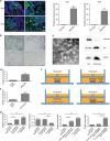
The mandible regulated tooth germ development through exosome-transferred ssc-mir-133b. a Apoptosis was measured with a TUNEL assay in E30 and E35 mandibles. Red arrowheads represent dental lamina. Scale bar = 200 μm. ***P < 0.001. b In situ hybridization of ssc-mir-133b in E30, E35, E40 and E50 mandibles. Scale bar = 200 μm. c Exosome characterization by transmission electron microscopy (left) and Western blotting (right). Scale bar = 50 nm. Calnexin is an endoplasmic reticulum marker and Alix and CD63 are exosome markers. d qPCR for ssc-mir-133b levels in E35 tooth germ and mandible. ***P < 0.001. e qPCR for ssc-mir-133b levels in E35 tooth germ and mandible exosomes. ***P < 0.001. f Transwell culture of tooth germ and mandible or mandible exosomes in vitro. g qPCR for ssc-mir-133b levels in tooth germ and mandible or mandible exosomes after Transwell culture. ***P < 0.001
Fig. 5
Fig. 5
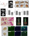
Mandible-derived ssc-mir-133b is indispensable in normal tooth formation. E40 premolars were cultured in vitro for 3 days with lentiviruses expressing neg-mir, miR-133b (left) and miR-133b + mcl-1 (right). They were then incubated subcutaneously in the back of nude mice for 12 weeks. a Tooth germs with lentiviruses expressing miR-133b failed to form, while tooth germs with lentiviruses expressing miR-133b + mcl-1 were readily observed 12 weeks after subcutaneous transplantation. b Morphological characteristics under a stereomicroscope. c 3D micro CT images of the negative control and Mcl-1 rescue groups. Tooth width, height of the teeth cups and tooth hardness were calculated for both groups. ***P < 0.001. d Haematoxylin and eosin staining of miR-133b + mcl-1 and neg-mir tooth germs. Scale bar = 200 μm. Dentin (d), dental pulp (DP), and enamel (e). e Status of tooth germ co-cultured with ma’ndible with (left side) and without GW4869 (right side). A red circle represents the transplantation area. f Morphology and amelogenin expression in tooth 12 weeks after co-culture with mandible with or without GW4869. Scale bar = 200 μm
Fig. 6
Fig. 6
Schematic of mandible exosomal ssc-mir-133b regulation in tooth morphogenesis. In tooth development, the mandible secretes exosomes to transfer crucial regulatory ssc-mir-133b to tooth germs. Targeting Mcl-1 and related endogenous mitochondrial apoptotic signals regulates the morphology of the developing tooth

Similar articles

Cited by

References

    1. Morriss-Kay GM, Sokolova N. Embryonic development and pattern formation. FASEB J. 1996;10:961–968. doi: 10.1096/fasebj.10.9.8801178. - DOI - PubMed
    1. Riobo NA, Lu K, Emerson CP., Jr Hedgehog signal transduction: signal integration and cross talk in development and cancer. Cell Cycle. 2006;5:1612–1615. doi: 10.4161/cc.5.15.3130. - DOI - PubMed
    1. Doroquez DB, Rebay I. Signal integration during development: mechanisms of EGFR and Notch pathway function and cross-talk. Crit. Rev. Biochem. Mol. Biol. 2006;41:339–385. doi: 10.1080/10409230600914344. - DOI - PubMed
    1. Francis-West P, Ladher R, Barlow A, Graveson A. Signalling interactions during facial development. Mech. Dev. 1998;75:3–28. doi: 10.1016/S0925-4773(98)00082-3. - DOI - PubMed
    1. Foster BL, et al. Development, disease, and regeneration of tissues in the dental-craniofacial complex. Biomed. Res. Int. 2013;2013:836871. doi: 10.1155/2013/836871. - DOI - PMC - PubMed