

Three-Dimensional Volumetric Segmentation of Pituitary Tumors: Assessment of Inter-rater Agreement and Comparison with Conventional Geometric Equations
- PMID: 30210975
- PMCID: PMC6133660
- DOI: 10.1055/s-0037-1618577
Three-Dimensional Volumetric Segmentation of Pituitary Tumors: Assessment of Inter-rater Agreement and Comparison with Conventional Geometric Equations
Abstract
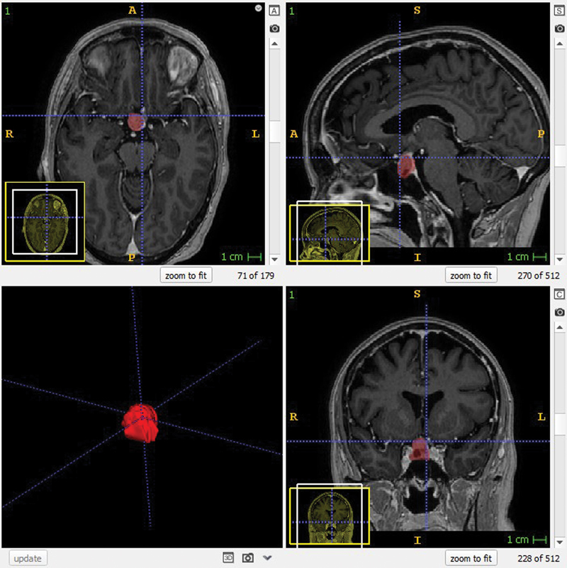
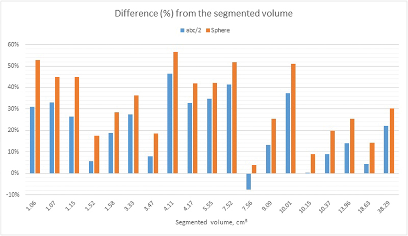
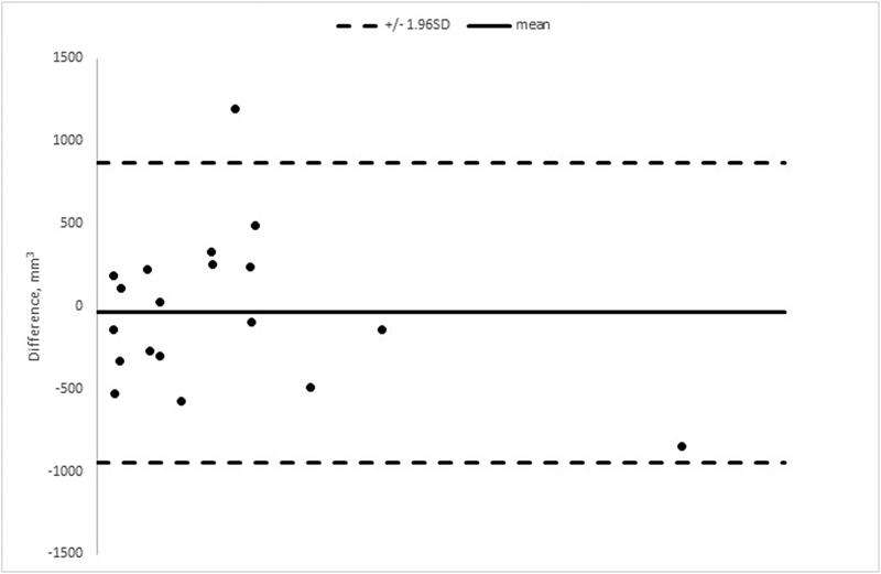
Background The assessment of pituitary tumor (PT) volume is important in the treatment and follow-up of patients with PT. Previously, PT volume estimation has been performed by conventional geometric equations (CGE) such as abc/2 (simplified ellipsoid volume equation) and 4πr 3 /3 (sphere), both presuming a symmetric tumor shape, which occurs uncommonly in patients with PT. In contrast, three-dimensional (3D) voxel-based software segmentation takes the irregular and asymmetric shapes that PTs often possess into account and might be a more accurate method for PT volume segmentation. The purpose of this study is twofold. (1) To compare 3D segmentation with CGE for PT volume estimation. (2) To assess inter-rater reliability in 3D segmentation of PTs. Methods Nineteen high-resolution (1mm slice thickness) T1-weighted MRI examinations of patients with PT were independently analyzed and manually segmented, using the software ITK-SNAP, by two certified neuroradiologists. Concurrently, the volumes of the PTs were estimated with abc/2 and 4πr 3 /3 by a clinician, and the results were compared with the corresponding segmented volumes. Results There was a significant decrease in PT volume attained from the segmentations compared with the calculations made with abc/2 ( p < 0.001, mean volume 18% higher than segmentation) and 4πr 3 /3 ( p < 0.001, mean volume 28% higher than segmentation). The intraclass correlation coefficient (ICC) for the two sets of segmented PTs was 0.99. Conclusion CGE ( abc/2 and 4πr 3 /3 ) significantly overestimates PT volume compared with 3D volumetric segmentation. The inter-rater agreement on manual 3D volumetric software segmentation is excellent.
Keywords: abc/2; pituitary tumor volume; segmentation; volumetric analysis.
Conflict of interest statement
Figures
Similar articles
-
Inter-rater agreement in glioma segmentations on longitudinal MRI.Neuroimage Clin. 2019;22:101727. doi: 10.1016/j.nicl.2019.101727. Epub 2019 Feb 22. Neuroimage Clin. 2019. PMID: 30825711 Free PMC article.
-
Glioblastoma Segmentation: Comparison of Three Different Software Packages.PLoS One. 2016 Oct 25;11(10):e0164891. doi: 10.1371/journal.pone.0164891. eCollection 2016. PLoS One. 2016. PMID: 27780224 Free PMC article.
-
Measuring glioma volumes: A comparison of linear measurement based formulae with the manual image segmentation technique.J Cancer Res Ther. 2016 Jan-Mar;12(1):161-8. doi: 10.4103/0973-1482.153999. J Cancer Res Ther. 2016. PMID: 27072231
-
Rapid Contour-based Segmentation for 18F-FDG PET Imaging of Lung Tumors by Using ITK-SNAP: Comparison to Expert-based Segmentation.Radiology. 2018 Jul;288(1):277-284. doi: 10.1148/radiol.2018171756. Epub 2018 Apr 3. Radiology. 2018. PMID: 29613842
-
Measuring pituitary tumor volume: a comparison of the simplified and non-simplified ellipsoid equation with the 3D planimetric volume assessment.Pituitary. 2023 Aug;26(4):383-392. doi: 10.1007/s11102-023-01317-4. Epub 2023 Apr 28. Pituitary. 2023. PMID: 37115292 Review.
Cited by
-
Mapping Pituitary Neuroendocrine Tumors: An Annotated MRI Dataset Profiling Tumor and Carotid Characteristics.Sci Data. 2025 Jan 15;12(1):80. doi: 10.1038/s41597-024-04218-8. Sci Data. 2025. PMID: 39814754 Free PMC article.
-
Fully automated grading of pituitary adenoma.Neuroimage Rep. 2025 Jan 29;5(1):100233. doi: 10.1016/j.ynirp.2025.100233. eCollection 2025 Mar. Neuroimage Rep. 2025. PMID: 40567882 Free PMC article.
-
Exploring the significance of tumor volume in endometrial cancer: Clinical pathological features, prognosis, and adjuvant therapies.Medicine (Baltimore). 2023 Dec 15;102(50):e36442. doi: 10.1097/MD.0000000000036442. Medicine (Baltimore). 2023. PMID: 38115321 Free PMC article.
-
Comparison of cerebrospinal fluid space between probable normal pressure hydrocephalus and Alzheimer's disease.Front Aging Neurosci. 2023 Aug 24;15:1241237. doi: 10.3389/fnagi.2023.1241237. eCollection 2023. Front Aging Neurosci. 2023. PMID: 37693646 Free PMC article.
-
In Vivo 3D MRI Measurement of Tumour Volume in an Orthotopic Mouse Model of Prostate Cancer.Cancer Control. 2019 Jan-Dec;26(1):1073274819846590. doi: 10.1177/1073274819846590. Cancer Control. 2019. PMID: 31032634 Free PMC article.
References
-
- Johannesen T B, Angell-Andersen E, Tretli S, Langmark F, Lote K. Trends in incidence of brain and central nervous system tumors in Norway, 1970-1999. Neuroepidemiology. 2004;23(03):101–109. - PubMed
-
- Materljan E, Materljan B, Sepcić J, Tuskan-Mohar L, Zamolo G, Erman-Baldini I. Epidemiology of central nervous system tumors in Labin area, Croatia, 1974-2001. Croat Med J. 2004;45(02):206–212. - PubMed
-
- Hardy J. New York, NY: Elsevier; 1973. Transsphenoidal surgery of hypersecreting pituitary tumors; pp. 179–194.
-
- Di Ieva A, Rotondo F, Syro L V, Cusimano M D, Kovacs K. Aggressive pituitary adenomas--diagnosis and emerging treatments. Nat Rev Endocrinol. 2014;10(07):423–435. - PubMed
LinkOut - more resources
Full Text Sources
Other Literature Sources
