. 2018 Oct;79(Suppl 4):S347-S355.
doi: 10.1055/s-0038-1654703. Epub 2018 May 25.

Less Invasive Modified Extradural Temporopolar Approach for Paraclinoid Lesions: Operative Technique and Surgical Results in 80 Consecutive Patients

Affiliations

Less Invasive Modified Extradural Temporopolar Approach for Paraclinoid Lesions: Operative Technique and Surgical Results in 80 Consecutive Patients

Naoki Otani et al. J Neurol Surg B Skull Base. 2018 Oct.

Abstract

Background Extradural temporopolar approach for paraclinoid lesions can provide extensive and early exposure of the anterior clinoid process, and complete mobilization and decompression of the optic nerve and internal carotid artery, which can prevent intraoperative neurovascular injury. The present study investigated the usefulness of our less invasive modified technique and discussed its operative nuances. Methods We retrospectively reviewed medical charts of 80 consecutive patients with neoplastic (21 patients) and vascular lesions (59 patients) who underwent the modified extradural temporopolar approach between September 2009 and March 2014. Results Preoperative visual acuity worsened in 4 patients (5.0%) and worsening of visual field function occurred in 10 patients (12.5%). Postoperative outcome was good recovery in 71 patients, moderate disability in 6, severe disability in 2, and death in 1 (due to reruptured aneurysm). No operation-related mortality occurred in the series. Conclusion Less invasive modified extradural temporopolar approach is safe and can be recommended for the surgical treatment of deeply located aneurysms and skull base tumors to reduce the risk of intraoperative optic neurovascular injury.

Keywords: anterior clinoid process; extradural temporopolar approach; paraclinoid aneurysm; paraclinoid tumors; skull base technique.


Figures

Fig. 1
Fig. 1
Schematic illustration of the operative techniques of modified extradural anterior clinoidectomy. Skeletonization of the superior orbital fissure (SOF) and only minimal dural incision between the SOF and foramen rotundum (FR) where no cranial nerves are present ( A , broken line) are performed, because the junction between the dura propria and the periosteal dura is invaginated at the SOF and carries the risk of injury to the cranial nerves. The dura propria should be carefully peeled from the SOF to preserve the sphenoparietal sinus (SPS) on the peeled dura propria until the anterior clinoid process (ACP) is entirely exposed epidurally ( B ). After removal of the ACP, the optic canal (OC) should be opened using a micro-punch. The clinoid segment (C3) of the internal carotid artery can be seen ( C ). Abbreviations: C3, clinoid segment of the internal carotid artery; MOB, meningo-orbital ban; II, optic nerve; III, oculomotor nerve; IV, trochlear nerve; V1, first division of the trigeminal nerve; V2, second division of the trigeminal nerve.
Fig. 2
Fig. 2
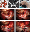
A 67-year-old female presented with mild headache and magnetic resonance imaging incidentally discovered paraclinoid carotid artery aneurysms on the right ( A , B ). The extradural temporopolar approach combined with orbitozygomatic osteotomy was performed ( CG ). The roof of the superior orbital fissure is skeletonized and opened to expose the junction between the dura propria of the temporal lobe and the periosteal dura, and the anterior clinoid process (ACP) is exposed epidurally ( D ). After removal of the ACP, the clinoid segment (C3) of the internal carotid artery can be seen ( E ). The falciform ligament is incised to mobilize the optic nerve (II). An additional incision is made across the distal dural ring to expose and identify the origin of the ophthalmic artery and to mobilize the internal carotid artery. The paraclinoid carotid artery aneurysm is dissected and clipped ( F , G ). The postoperative course was uneventful. Abbreviations: OC, optic canal; C2, ophthalmic segment of the internal carotid artery; C3, clinoid segment of the internal carotid artery; II, optic nerve; V1, first division of the trigeminal nerve; V2, second division of the trigeminal nerve.
Fig. 3
Fig. 3
A 43-year-old male presented with right progressive visual disturbance. Three-dimensional computed tomography angiography showed an abnormal lesion located at the tuberculum sellae ( A ). The extradural temporopolar approach with standard frontotemporal craniotomy was performed. The foramen rotundum (FR) and superior orbital fissure (SOF) are exposed ( B ). The roof of the SOF is skeletonized and opened to expose the junction between the dura propria of the temporal lobe and the periosteal dura. The anterior clinoid process (ACP) is exposed epidurally ( C ). After removal of the ACP, the falciform ligament (FL) is incised ( D ) to mobilize the optic nerve (II). Gross total removal was performed. Postoperative course was uneventful and visual examination showed normalized visual disturbance. Abbreviations: IC, internal carotid artery; V1, first division of the trigeminal nerve; V2, second division of the trigeminal nerve.

References

    1. Guidetti B, La Torre E. Management of carotid-ophthalmic aneurysms. J Neurosurg. 1975;42(04):438–442. - PubMed
    1. Sundt T M, Jr, Piepgras D G. Surgical approach to giant intracranial aneurysms. Operative experience with 80 cases. J Neurosurg. 1979;51(06):731–742. - PubMed
    1. Day J D, Giannotta S L, Fukushima T. Extradural temporopolar approach to lesions of the upper basilar artery and infrachiasmatic region. J Neurosurg. 1994;81(02):230–235. - PubMed
    1. Lee J H, Jeun S S, Evans J, Kosmorsky G.Surgical management of clinoidal meningiomas Neurosurgery 200148051012–1019., discussion 1019–1021 - PubMed
    1. Evans J J, Hwang Y S, Lee J H.Pre- versus post-anterior clinoidectomy measurements of the optic nerve, internal carotid artery, and opticocarotid triangle: a cadaveric morphometric study Neurosurgery 200046041018–1021., discussion 1021–1023 - PubMed