Review
. 2018 Aug;8(7):673-693.
doi: 10.21037/qims.2018.08.08.

Comprehensive genotype-phenotype correlation in lissencephaly

Affiliations
Review

Comprehensive genotype-phenotype correlation in lissencephaly

Ai Peng Tan et al. Quant Imaging Med Surg. 2018 Aug.

Abstract

Malformations of cortical development (MCD) are a heterogenous group of disorders with diverse genotypic and phenotypic variations. Lissencephaly is a subtype of MCD caused by defect in neuronal migration, which occurs between 12 and 24 weeks of gestation. The continuous advancement in the field of molecular genetics in the last decade has led to identification of at least 19 lissencephaly-related genes, most of which are related to microtubule structural proteins (tubulin) or microtubule-associated proteins (MAPs). The aim of this review article is to bring together current knowledge of gene mutations associated with lissencephaly and to provide a comprehensive genotype-phenotype correlation. Illustrative cases will be presented to facilitate the understanding of the described genotype-phenotype correlation.

Keywords: LIS1; Lissencephaly (LIS); doublecortin (DCX); tubulinopathy.


Conflict of interest statement

Conflicts of Interest: The authors have no conflicts of interest to declare.

Figures

Figure 1
Figure 1
Lissencephaly (LIS)-associated genes, many of which are related to microtubule structural proteins (tubulin) or microtubule-associated proteins (MAPs).
Figure 2
Figure 2
Radiological characterisation of LIS. Cerebellar hypoplasia/dysplasia, dysmorphic basal ganglia, thin or absent corpus callosum, ventricular dilation, and abnormalities of the hippocampus and brainstem are useful diagnostic indicators of tubulin gene involvement. Severe cerebellar/brainstem hypoplasia is also seen in association with RELN/VLDLR and NDE1 mutations. In the presence of intracranial calcifications and ventriculomegaly, congenital CMV/TORCH infection needs to be excluded. LIS, lissencephaly.
Figure 3
Figure 3
Roles of LIS-associated genes in association with microtubule function and neuronal migration/organization. (I) Mutations in tubulin isotypes disrupt the formation of normal heterodimers of α- and β-tubulin polypeptides, affecting the structural stability and function of microtubules; (II) kinesins and (III) dyneins are microtubule motor proteins that power directional movement along microtubules. Lissencephaly (LIS)-related genes that encode these motor proteins include KIF2A, KIF5C and DYNC1H1; (IV) the LIS1 gene encodes the LIS1 protein which is an adaptor for the microtubule-motor dynein that allows dynein to remain attached to microtubules for longer periods of time; (V) the mammalian NudE homologues (NDE1 and NDEL1) are required for the targeting of LIS1 to the dynein complex. It also releases the blocking effect of LIS1 on cytoplasmic dynein; (VI) DCX protein binds directly to microtubules to stabilize and promote polymerization, facilitates the formation of the microtubule cage around the nucleus, as well as stabilizes microtubules in the leading process of the migrating neuron; (VII) the Reelin signalling pathway aids in the regulation of neuronal migration and positioning, generating the “inside first-outside last” configuration of the 6-layer cortex; (VIII) CDK5 regulates the binding and assembly of LIS1-dynein complex as well as the direct binding of DCX to microtubules; (IX) the ARX gene encodes the ARX protein (a transcription factor) which regulates genes that play crucial roles in tangential migration of GABAergic interneurons into the cortical plate; (X) ACTB and ACTG1 encode β- and γ-actin. The interaction between microtubules and cytoplasmic actin plays an important role in neuronal migration. DCX protein allows “cross-talk” between microtubules and cytoplasmic actins; (XI) the CRADD gene is a recently discovered LIS-associated gene. Its role in neuronal migration and hence development of LIS is still unclear and needs further investigation. MCD, malformations of cortical development.
Figure 4
Figure 4
Classical lissencephaly in a patient with heterozygous deletion in the LIS1 gene. (A,B) Axial T2-weighted images demonstrate the presence of posterior predominant LIS with a thick cortex. Mild ventriculomegaly is noted; (C) sagittal T1-weighted and (D) coronal T2-weighted images show normal appearances of the cerebellum and brainstem. Partial agenesis of the corpus callosum is also noted.
Figure 5
Figure 5
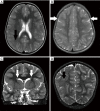
Posterior predominant pachygyria with underlying SBH in a patient with mosaic mutations of LIS1. (A) Axial CT image shows bilateral pachygyria with mild ventriculomegaly; (B) axial T2-weighted and (D,E) coronal T2-weighted images demonstrate the presence of bilateral pachygyria, more severe posteriorly. An underlying band of heterotopic grey matter (white arrows) is also present posteriorly; (C) sagittal T1-weighted image shows normal appearances of the cerebellum, brainstem and corpus callosum. SBH, subcortical band heterotopia.
Figure 6
Figure 6
DCX-related lissencephaly due to a missense mutation. (A-C) Axial T2-weighted images show bilateral pachygyria with thick cortex (involving predominantly the frontoparietal lobes) and moderate ventriculomegaly. The basal ganglia is normal in appearance; (D) sagittal T1-weighted image shows severe hypoplasia of the corpus callosum (white arrow). The brainstem and vermis are normal; (E,F) coronal FLAIR images confirms the presence of morphologically normal cerebellar hemispheres and abnormal orientation of the hippocampi.
Figure 7
Figure 7
DCX-related SBH in a female patient. (A,B) Axial and (C) coronal T2-weighted images show bilateral SBH with ventriculomegaly and prominent perivascular spaces. The basal ganglia and cerebellum are normal; (D) sagittal T1-weighted image shows mild hypoplasia of the posterior corpus callosum with loss of the normal bulbous appearance of the splenium (white arrow). SBH, subcortical band heterotopia.
Figure 8
Figure 8
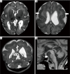
TUBA1A-related lissencephaly. (A-C) Axial T2-weighted images demonstrate bilateral perisylvian pachygyria-polymicrogyria with mild ventriculomegaly and signal abnormalities within the cerebral white matter. There is also absence of the anterior limb of the internal capsules with fusion of the caudate head and lentiform nucleus bilaterally (black arrow); (D) coronal T1-weighted image shows tiny cysts within the left cerebellar hemisphere (white arrow); (E) axial T2-weighted MRI image shows bilateral intra-ocular lens replacement due to congenital cataracts; (F) sagittal T1-weighted image shows partial agenesis of the corpus callosum and mild hypoplasia of the brainstem.
Figure 9
Figure 9
TUBB2B-related lissencephaly. (A,B) Axial T2-weighted images show anterior predominant pachygyria with thin cortex and underlying SBH (white arrows). There is also absence of the anterior limb of the internal capsule with fusion of the caudate head and lentiform nucleus bilaterally. The ventricles are asymmetrically enlarged; (C) coronal T2-weighted and (D) sagittal T1-weighted images show pontocerebellar hypoplasia as well as mild hypoplasia of the corpus callosum. SBH, subcortical band heterotopia.
Figure 10
Figure 10
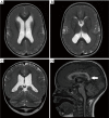
TUBA1A-related lissencephaly. (A-C) Axial T2-weighted images show bilateral central pachygyria-polymicrogyria with subcortical band of heterotopic grey matter (thin white arrow in A). There is also fusion of the caudate head and lentiform nucleus bilaterally due to absence of the anterior limb of the internal capsules. Complete corpus callosal agenesis is noted with colpocephaly; (D,E) axial T2-weighted image across the posterior fossa shows hypoplasia and abnormal shape of the pons. The cerebellar vermis is deficient with dysplasia of the cerebellar hemispheres and the presence of tiny cerebellar hemispheric cysts (black arrows in E); (F) 3D CT image shows severe microcephaly.
Figure 11
Figure 11
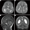
X-linked lissencephaly with abnormal genitalia (XLAG). This child presented with intractable seizures soon after birth and was noted to have micropenis and bilateral undescended testis on examination. (A) Axial T2-weighted; (B) coronal T2-weighted and (C) sagittal T1-weighted images show a simplified gyral pattern and perisylvian pachygyria (white arrows in A), along with hypoplasia of the brainstem, vermis and cerebellar hemispheres. There is complete agenesis of the corpus callosum with an interhemispheric cyst (*). Of note is also the presence of microcephaly.
Figure 12
Figure 12
Actin-isoform gene-related lissencephaly. (A,B) Axial T2-weighted and (C,D) coronal T2-weighted images show subtle anterior pachygyria/simplified gyral pattern (white arrows in B) with posterior SBH (black arrows in A and D). Bilateral frontal periventricular nodular heterotopia (white arrows in C) is also observed. SBH, subcortical band heterotopia.
Figure 13
Figure 13
Actin-isoform gene-related LIS. (A,B) Axial T2-weighted and (C-E) coronal T2-weighted images demonstrate frontal predominant pachygyria merging with posterior subcortical band heterotopia. Mild ventriculomegaly is also noted. The cerebellar hemispheres are normal. LIS, lissencephaly.

References

    1. des Portes V, Francis F, Pinard JM, Desguerre I, Moutard ML, Snoeck I, Meiners LC, Capron F, Cusmai R, Ricci S, Motte J, Echenne B, Ponsot G, Dulac O, Chelly J, Beldjord C. Doublecortin is the major gene causing X-linked subcortical laminar heterotopia (SCLH). Hum Mol Genet 1998;7:1063-70. 10.1093/hmg/7.7.1063 - DOI - PubMed
    1. Gleeson JG, Allen KM, Fox JW, Lamperti ED, Berkovic S, Scheffer I, Cooper EC, Dobyns WB, Minnerath SR, Ross ME, Walsh CA. Doublecortin, a brain-specific gene mutated in human X-linked lissencephaly and double cortex syndrome, encodes a putative signaling protein. Cell 1998;92:63-72. 10.1016/S0092-8674(00)80899-5 - DOI - PubMed
    1. Reiner O, Carrozzo R, Shen Y, Wehnert M, Faustinella F, Dobyns WB, Caskey CT, Ledbetter DH. Isolation of a Miller-Dieker lissencephaly gene containing G protein beta-subunit-like repeats. Nature 1993;364:717-21. 10.1038/364717a0 - DOI - PubMed
    1. Di Donato N, Chiari S, Mirzaa GM, Aldinger K, Parrini E, Olds C, Barkovich AJ, Guerrini R, Dobyns WB. Lissencephaly: Expanded imaging and clinical classification. Am J Med Genet A 2017;173:1473-88. 10.1002/ajmg.a.38245 - DOI - PMC - PubMed
    1. Abdollahi MR, Morrison E, Sirey T, Molnar Z, Hayward BE, Carr IM, Springell K, Woods CG, Ahmed M, Hattingh L, Corry P, Pilz DT, Stoodley N, Crow Y, Taylor GR, Bonthron DT, Sheridan E. Mutation of the variant alpha-tubulin TUBA8 results in polymicrogyria with optic nerve hypoplasia. Am J Hum Genet 2009;85:737-44. 10.1016/j.ajhg.2009.10.007 - DOI - PMC - PubMed

LinkOut - more resources