Polyethylene Oxide and Silicon-Substituted Hydroxyapatite Composite: A Biomaterial for Hard Tissue Engineering in Orthopedic and Spine Surgery
- PMID: 30211130
- PMCID: PMC6124219
- DOI: 10.4103/abr.abr_206_17
Polyethylene Oxide and Silicon-Substituted Hydroxyapatite Composite: A Biomaterial for Hard Tissue Engineering in Orthopedic and Spine Surgery
Abstract
Background: Tissue engineering and biomaterials have made it possible to innovate bone treatments for orthopedic and spine problems. The aim of this study is to develop a novel polyethylene oxide (PEO)/silicon-substituted hydroxyapatite (Si-HA) composite to be used as a scaffold for hard tissue engineering in orthopedic and spine procedures.
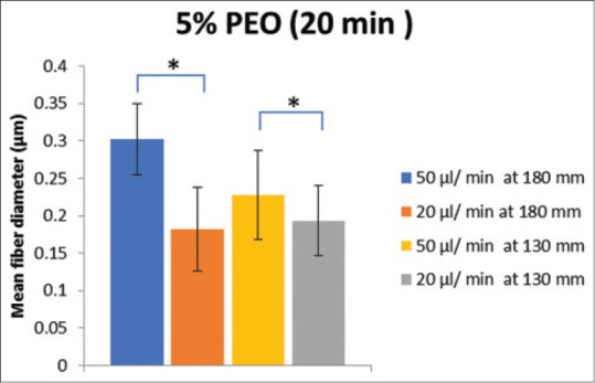
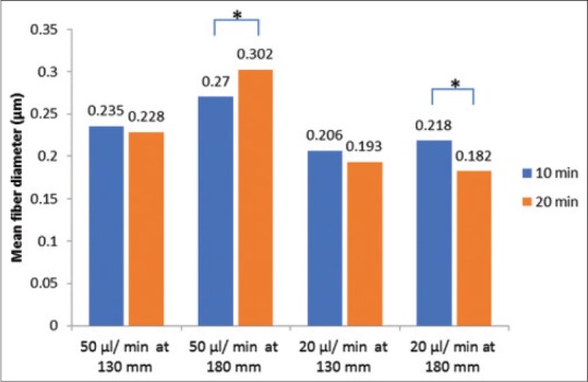
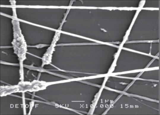
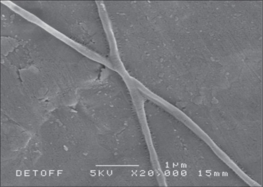
Materials and methods: The composite was fabricated through the electrospinning technique. The applied voltage (5 kV) and PEO concentration (5%) were fixed. Processing parameters such as the flow rates (20 μl/min and 50 μl/min), distances from capillary tube to the collector (130 mm and 180 mm), spinning time (10 min and 20 min), and concentration of Si-HA (0.2% and 0.6%) were explored to find the optimum conditions to produce fine composite fibers.
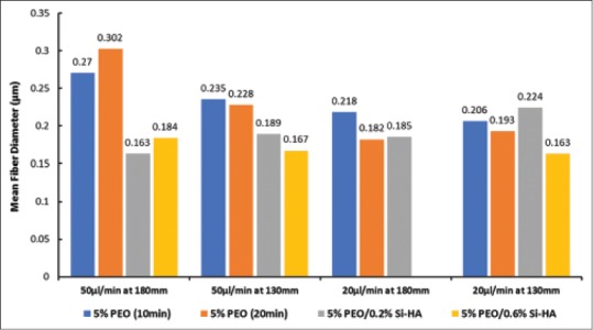
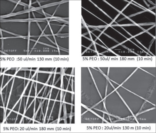
Results: Scanning electron microscope images showed that 5% PEO, 5% PEO/0.2% Si-HA, and 5% PEO/0.6% Si-HA fibers were successively produced. Flow rates and working distances showed significant influence on the morphology of the polymeric and composite fibers. A high flow rate (50 μl/min) and a larger working distance (180 mm) resulted in larger fibers. The comparison between the mean fiber diameter of 5% PEO/0.2% Si-HA and 5% PEO/0.6% Si-HA showed to be significantly different. As the Si-HA concentration increased, certain fibers were having particles of Si-HA that were not properly integrated into the polymer matrix.
Conclusions: Synthesis of a novel biomaterial for hard tissue scaffold through electrospinning was successful. In general, PEO/Si-HA fibers produced have the desired characteristics to mimic the extracellular matrix of bone.
Keywords: Biomaterial; hard tissue engineering; neurosurgery; orthopedics; polyethylene oxide; silicon-substituted hydroxyapatite; spine surgery.
Conflict of interest statement
There are no conflicts of interest.
Figures


References
-
- National Osteoporosis Society. A Strategy to Reduce the Impact of Osteoporosis and Fragility Fractures in England. 2009. [Last accessed on 2018 Jun 20]. Available from: http://www.nos.org.uk/document.doc?id=491 .
-
- Cooper C, Campion G, Melton LJ., 3rd Hip fractures in the elderly: A world-wide projection. Osteoporos Int. 1992;2:285–9. - PubMed
-
- Huiskes R, Weinans H, van Rietbergen B. The relationship between stress shielding and bone resorption around total hip stems and the effects of flexible materials. Clin Orthop Relat Res. 1992;274:124–34. - PubMed
-
- Hench LL, Polak JM. Third-generation biomedical materials. Science. 2002;295:1014–7. - PubMed
-
- Archibeck MJ, Berger RA, Jacobs JJ, Quigley LR, Gitelis S, Rosenberg AG, et al. Second-generation cementless total hip arthroplasty. Eight to eleven-year results. J Bone Joint Surg Am. 2001;83-A:1666–73. - PubMed
LinkOut - more resources
Full Text Sources
Other Literature Sources