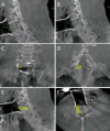
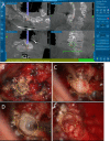
Augmented Reality Visualization-guided Microscopic Spine Surgery: Transvertebral Anterior Cervical Foraminotomy and Posterior Foraminotomy
- PMID: 30211385
- PMCID: PMC6132325
- DOI: 10.5435/JAAOSGlobal-D-17-00008
Augmented Reality Visualization-guided Microscopic Spine Surgery: Transvertebral Anterior Cervical Foraminotomy and Posterior Foraminotomy
Abstract
Objective: We describe intraoperative augmented reality (AR) imaging to obtain a microscopic view in spine keyhole surgery.
Background: Minimally invasive keyhole surgery has been developed even for spine surgery, including transvertebral anterior cervical foraminotomy and posterior cervical laminoforaminotomy. These methods are complex and require a skillful technique. Therefore, inexperienced surgeons hesitate to perform keyhole surgeries. The technology used in surgery is rapidly advancing, including intraoperative imaging devices that have enabled AR imaging and facilitated complicated surgeries in many fields. However, data are not currently available on the use of AR imaging in spine surgery. The purpose of this article was to introduce the utility of AR for spine surgery.
Methods: We performed O-arm intraoperative imaging to create an augmented imaging model in navigation systems. Navigation data were linked to a microscope to merge the live view and AR. Augmented reality imaging shows the model plan in the real-world surgical field. We used this novel method in patients who underwent both keyhole surgeries.
Results: We successfully performed both surgeries using the AR visualization guide.
Conclusions: The AR navigation system facilitates complicated keyhole surgeries in patients who undergo spine surgery.
Study design: Technical report.
Conflict of interest statement
None of the following authors or any immediate family member has received anything of value from or has stock or stock options held in a commercial company or institution related directly or indirectly to the subject of this article: Dr. Umebayashi, Dr. Yamamoto, Dr. Nakajima, Dr. Fukaya, and Dr. Hara. Dr. Umebayashi designed research. Dr. Umebayashi, Dr. Yamamoto, Dr. Nakajima, Dr. Fukaya, and Dr. Hara performed research. Dr. Umebayashi analyzed the data and wrote the article. The device(s)/drug(s) is approved by the FDA or by the corresponding national agency for this indication.
Figures


Similar articles
-
Minimally Invasive Full-Endoscopic Posterior Cervical Foraminotomy Assisted by O-Arm-Based Navigation.Pain Physician. 2018 May;21(3):E215-E223. Pain Physician. 2018. PMID: 29871377
-
Augmented and virtual reality in spine surgery, current applications and future potentials.Spine J. 2021 Oct;21(10):1617-1625. doi: 10.1016/j.spinee.2021.03.018. Epub 2021 Mar 25. Spine J. 2021. PMID: 33774210 Review.
-
Virtual reality in spinal endoscopy: a paradigm shift in education to support spine surgeons.J Spine Surg. 2020 Jan;6(Suppl 1):S208-S223. doi: 10.21037/jss.2019.11.16. J Spine Surg. 2020. PMID: 32195429 Free PMC article.
-
Augmented Reality Surgical Navigation in Minimally Invasive Spine Surgery: A Preclinical Study.Bioengineering (Basel). 2023 Sep 18;10(9):1094. doi: 10.3390/bioengineering10091094. Bioengineering (Basel). 2023. PMID: 37760196 Free PMC article.
-
Cervical Spine Surgery: Arthroplasty Versus Fusion Versus Posterior Foraminotomy.Neurosurg Clin N Am. 2021 Oct;32(4):483-492. doi: 10.1016/j.nec.2021.05.005. Neurosurg Clin N Am. 2021. PMID: 34538474 Review.
Cited by
-
The utility of virtual reality and augmented reality in spine surgery.Ann Transl Med. 2019 Sep;7(Suppl 5):S171. doi: 10.21037/atm.2019.06.38. Ann Transl Med. 2019. PMID: 31624737 Free PMC article. Review.
-
XR (Extended Reality: Virtual Reality, Augmented Reality, Mixed Reality) Technology in Spine Medicine: Status Quo and Quo Vadis.J Clin Med. 2022 Jan 17;11(2):470. doi: 10.3390/jcm11020470. J Clin Med. 2022. PMID: 35054164 Free PMC article. Review.
-
Robotic Spine Surgery and Augmented Reality Systems: A State of the Art.Neurospine. 2020 Mar;17(1):88-100. doi: 10.14245/ns.2040060.030. Epub 2020 Mar 31. Neurospine. 2020. PMID: 32252158 Free PMC article.
-
Augmented and virtual reality in spine surgery.J Orthop. 2023 Jul 18;43:30-35. doi: 10.1016/j.jor.2023.07.018. eCollection 2023 Sep. J Orthop. 2023. PMID: 37555206 Free PMC article.
-
Advancements and Challenges in Computer-Assisted Navigation for Cervical Spine Surgery: A Comprehensive Review of Perioperative Integration, Complications, and Emerging Technologies.Global Spine J. 2025 Apr 4:21925682251329340. doi: 10.1177/21925682251329340. Online ahead of print. Global Spine J. 2025. PMID: 40183132 Free PMC article. Review.
References
-
- Shuhaiber JH: Augmented reality in surgery. Arch Surg 2004;139:170-174. - PubMed
-
- Graham M, Zook M, Boulton A: Augmented reality in urban places: Contested content and the duplicity of code. Trans Inst Br Geogr 2012;38:464-479.
-
- Cabrilo I, Bijlenga P, Schaller K: Augmented reality in the surgery of cerebral aneurysms: A technical report. Neurosurgery 2014;10(suppl 2):252-260; discussion 260-261. - PubMed
-
- Cabrilo I, Bijlenga P, Schaller K: Augmented reality in the surgery of cerebral arteriovenous malformations: Technique assessment and considerations. Acta Neurochir (Wien) 2014;156:1769-1774. - PubMed
Publication types
LinkOut - more resources
Full Text Sources
Other Literature Sources
Research Materials

