

Acute Paravertebral Compartment Syndrome: Follow-up and Literature Review
- PMID: 30211387
- PMCID: PMC6132322
- DOI: 10.5435/JAAOSGlobal-D-17-00063
Acute Paravertebral Compartment Syndrome: Follow-up and Literature Review
Abstract
Objective: To report on a patient with acute paravertebral and posterior thigh compartment syndrome after vigorous exercise.
Background: Paravertebral compartment syndrome (PCS) is a rare clinical entity, typically occurring in male athletes after heavy exertion and weightlifting.
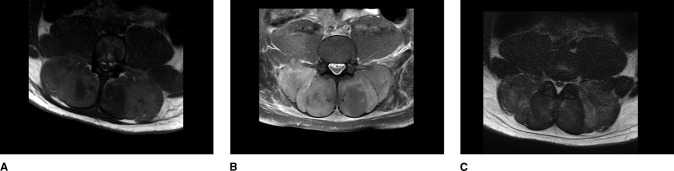
Case: A 25-year-old man presented with back pain and hematuria hours after back-specific weightlifting. Clinical examination, laboratory markers, MRI, and elevated intracompartmental pressure measurements supported the diagnosis of bilateral paravertebral and posterior thigh compartment syndrome. The patient underwent paravertebral decompression via the Wiltse approach with immediate postoperative relief. He is doing well at 1 year, with recovery of lumbar extension strength, although MRI demonstrates moderate fatty replacement of paravertebral musculature.
Conclusions: Although rare, early recognition of PCS and timely decompression can limit myonecrosis. Paravertebral compartment syndrome should be considered in the differential for athletic individuals with acute onset back pain.
Study design: A case report and review of literature.
Conflict of interest statement
None of the following authors or any immediate family member has received anything of value from or has stock or stock options held in a commercial company or institution related directly or indirectly to the subject of this article: Dr. Roe, Dr. Chen, and Dr. Cho.
Figures


References
-
- Kitajima I, Tachibana S, Hirota Y, Nakamichi K: Acute paraspinal muscle compartment syndrome treated with surgical decompression: A case report. Am J Sports Med 30:283-285. - PubMed
-
- Schreiber VM, Ward WT: Exercise-induced pediatric lumbar paravertebral compartment syndrome: A case report. J Pediatr Orthop 2015;35:e49-e51. - PubMed
-
- DiFazio FA, Barth RA, Frymoyer JW: Acute lumbar paraspinal compartment syndrome: A case report. J Bone Joint Surg Am 1991;73:1101-1103. - PubMed
-
- Rha EY, Kim DH, Yoo G: Acute exertional lumbar paraspinal compartment syndrome treated with fasciotomy and dermatotraction: Case report. J Plast Reconstr Aesthet Surg 2014;67:425-426. - PubMed
Publication types
LinkOut - more resources
Full Text Sources
Other Literature Sources
