. 2019 Jan 15;199(2):199-210.
doi: 10.1164/rccm.201707-1370OC.

Toll-like Receptor 3 Is a Therapeutic Target for Pulmonary Hypertension

Affiliations

Toll-like Receptor 3 Is a Therapeutic Target for Pulmonary Hypertension

Daniela Farkas et al. Am J Respir Crit Care Med. .

Abstract

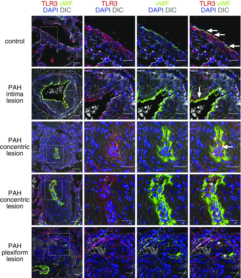
Rationale: Pulmonary arterial hypertension (PAH) is characterized by vascular cell proliferation and endothelial cell apoptosis. TLR3 (Toll-like receptor 3) is a receptor for double-stranded RNA and has been recently implicated in vascular protection.

Objectives: To study the expression and role of TLR3 in PAH and to determine whether a TLR3 agonist reduces pulmonary hypertension in preclinical models.

Methods: Lung tissue and endothelial cells from patients with PAH were investigated by polymerase chain reaction, immunofluorescence, and apoptosis assays. TLR3-/- and TLR3+/+ mice were exposed to chronic hypoxia and SU5416. Chronic hypoxia or chronic hypoxia/SU5416 rats were treated with the TLR3 agonist polyinosinic/polycytidylic acid (Poly[I:C]).

Measurements and main results: TLR3 expression was reduced in PAH patient lung tissue and endothelial cells, and TLR3-/- mice exhibited more severe pulmonary hypertension following exposure to chronic hypoxia/SU5416. TLR3 knockdown promoted double-stranded RNA signaling via other intracellular RNA receptors in endothelial cells. This was associated with greater susceptibility to apoptosis, a known driver of pulmonary vascular remodeling. Poly(I:C) increased TLR3 expression via IL-10 in rat endothelial cells. In vivo, high-dose Poly(I:C) reduced pulmonary hypertension in both rat models in proof-of-principle experiments. In addition, Poly(I:C) also reduced right ventricular failure in established pulmonary hypertension.

Conclusions: Our work identifies a novel role for TLR3 in PAH based on the findings that reduced expression of TLR3 contributes to endothelial apoptosis and pulmonary vascular remodeling.

Keywords: apoptosis; double-stranded RNA; endothelial cell; pulmonary hypertension; toll-like receptor 3.


Figures

Figure 1.
Figure 1.
Reduced endothelial TLR3 (Toll-like receptor 3) in lung vascular lesions of patients with pulmonary arterial hypertension. Double immunofluorescence shows strong immunostaining of TLR3 in von Willebrand factor–positive endothelium in pulmonary artery of control subject (arrows). TLR3 staining is partially lost in endothelium of pulmonary arterial hypertension pulmonary arteries with increased smooth muscle layer and intima thickening. The arrows indicate endothelium with preserved TLR3 expression, whereas asterisks indicate endothelium that is TLR3 deficient. In concentric and plexiform lesions, TLR3 staining is largely lost in von Willebrand factor–positive endothelium (asterisks). Scale bars: 50 μm (overview on the left), 25 μm (higher detail images). The dashed boxes in the overview images on the left indicate the area that is shown in more detail on the right. Nuclear counterstaining with DAPI. DIC = differential interference contrast; PAH = pulmonary arterial hypertension; vWF = von Willebrand factor.
Figure 2.
Figure 2.
Reduced TLR3 (Toll-like receptor 3) expression in human pulmonary arterial hypertension (PAH) and experimental severe pulmonary hypertension. (A) Lower expression of TLR3 mRNA in pulmonary artery endothelial cells (PAEC) (A, n = 4 control subjects vs. n = 3 PAH) from human patients with PAH as compared with control subjects without pulmonary vascular disease. (B) Reduced TLR3 protein expression in PAEC from patients with PAH by Western blot. β-Actin was used to ensure equal loading of lanes. (C) Semiquantitative densitometry of TLR3 versus β-actin confirms reduced TLR3 protein expression in PAEC from patients with PAH. n = 4 different cell lines per group. (D) Reduced mRNA expression of TLR3 in the lung tissue from patients with PAH (n = 5 control subjects vs. n = 6 PAH). Representative Western blot (E) and semiquantitative densitometry (F) show reduction of TLR3 protein expression in lung tissue protein lysate from chronic hypoxia and SU5416 (cHx/Su) rats at Day 21. β-Actin was used as loading control. n = 3 per group. (G) Representative immunohistochemistry images for TLR3 show less TLR3+ cells (arrows) in endothelial/intima cells in cHx and cHx/Su rats. Counterstaining: Mayer's hematoxylin. Scale bars: 20 μm. (H) Quantification of TLR3+ intima cells in pulmonary arteries of cHx and cHx/Su rats. n = 3 per group. (I) Representative images of von Willebrand factor and α-smooth muscle actin immunohistochemistry show increased pulmonary artery muscularization in TLR3−/− mice exposed to the cHx/Su protocol compared with TLR3+/+ wild-type mice. In contrast, no substantial change occurred in TLR3−/− mice housed in normoxia. Counterstaining with hematoxylin. Scale bars: 20 μm. TLR3−/− mice have higher right ventricular systolic pressure (J), media wall thickness (K), and fraction of muscularized pulmonary arteries (L) than TLR3 wild-type mice when exposed to the cHx/Su protocol but not when exposed to normoxia. n = 6–12 (J), n = 3–7 (K and L). Bars: mean + SEM, scatter plots indicate mean ± SEM. *P < 0.05, **P < 0.01, ***P < 0.0001 (A, C, and D: Student’s t test; F and H: one-way ANOVA; J–L: two-way ANOVA). MWT = media wall thickness; RVSP = right ventricular systolic pressure; SMA = smooth muscle actin; vWF = von Willebrand factor; WT = wild type.
Figure 3.
Figure 3.
Endothelial TLR3 (Toll-like receptor 3) deficiency promotes apoptosis and impairs migration in vitro. (A) Representative dot plots show increased fraction of annexin V+ (AV+) 7-AAD (apoptotic) cells in pulmonary artery endothelial cells (PAEC) from patients with pulmonary arterial hypertension. (B) Quantification of AV+ 7-AAD PAEC according to group (n = 6 each group). (C) Representative dot plots indicate increased fraction of AV+ 7-AAD CD117+ rat lung EC after short hairpin RNA–mediated knockdown of TLR3. (D) Quantification of AV+ 7-AAD CD117+ rat lung EC 72 hours after adenovirus-mediated overexpression of scrambled [scrm] short hairpin RNA or shTlr3 (n = 3 per group). (E) Gene knockdown was confirmed by qRT-PCR of rat Tlr3 gene mRNA expression 72 hours after the beginning of the transfection. (F) Representative Western blot shows reduction of TLR3 expression in control PAEC 48 hours after overexpression of cas9 and TLR3 sgRNA (or scrm sgRNA, as control). β-actin was used as loading control. (G) Semiquantitative densitometry (n = 3). (H) Immunofluorescence staining for TLR3 shows strong TLR3 expression in scrm CRISPR PAEC, but loss of TLR3 expression in most TLR3 CRISPR PAEC after 72 hours. The images on the top show an overview (scale bar: 50 μm) and the images on the bottom show cells in more detail, with the scrm cells exhibiting a typical cytoplasmic TLR3 staining pattern. Arrows indicate cells that retained TLR3 expression. (I) qRT-PCR shows mRNA expression of DDX58 (RIG-I) and IFIH1 (MDA-5) in TLR3 and scrm CRISPR PAEC. (J) Representative dot plots show increased fraction of AV+/7-AAD PAEC following serum starvation (basal endothelial growth medium) and TLR3 knockdown. (K) Quantification of AV+/7-AAD PAEC. n = 9 per group. (L) Representative differential interference contrast images of gap closure assay of PAEC after TLR3-targeted or scrm CRISPR. Cells from both groups were treated with vehicle or 100 μM Z-Asp-CH2-DCB. The images show the damage-free gap at 0 and 15 hours. The borders of the gaps are indicated by yellow dotted lines. Scale bar: 100 μm. (M) Quantification of percent wound closure after 15 hours (n = 9 per group). Bars: mean + SEM. *P < 0.05, **P < 0.01, ***P < 0.001, ****P < 0.0001 (B, D, E, G, and I: Student’s t test; K and M: two-way ANOVA). CRISPR = clustered, regularly interspaced short palindromic repeats; EGM = endothelial growth medium; PAH = pulmonary arterial hypertension; shscrm = scrambled short hairpin RNA; veh = vehicle.
Figure 4.
Figure 4.
TLR3 (Toll-like receptor 3) deficiency channels endothelial dsRNA signaling through alternate RNA receptors RIG-I and MDA-5 and promotes IL-10 expression in rat lung CD117+ endothelial cells (EC). (A) Representative optical sections of images acquired by confocal microscopy show that rhodamine-labeled polyinosinic/polycytidylic acid (Poly[I:C]) (25 μg/ml) localized to TLR3, RIG-I, and MDA-5 in shscrm-expressing CD117+ EC with normal TLR3 expression. By contrast, in shTlr3-expressing cells with reduced TLR3 level, Poly(I:C) mainly interacted with RIG-I and MDA-5. Arrows: colocalization of Poly(I:C) and respective receptor. Scale bars: 10 μm. The lower row shows the areas indicated by dashed boxes in the upper row in more detail. Counterstaining with DAPI. (B–E) Changes in the Poly(I:C)–induced mRNA expression of genes regulating inflammation and vasotonus/remodeling between shscrm- and shTlr3 EC: Il10 (B), Edn1 (Endothelin-1, C), Cxcl10 (CXCL10, D), and Il6 (E). (F and G) Whereas a low dose of Poly(I:C) (0.1 μg/ml) fails to induce Il10 (F) or Tlr3 (G) mRNA expression in CD117+ EC, high concentration of Poly(I:C) (50 μg/ml) strongly elevates expression of Il10 and Tlr3. (H and I) Poly(I:C)–induced elevation (25 μg/ml) of Tlr3 expression depends on IL-10, because treatment with a neutralizing anti-IL-10 antibody (ab) abolishes Poly(I:C)–induced upregulation of Tlr3 (H) but enhances Poly(I:C)–induced Cxcl10 upregulation (I). (J) Representative Western blots from nuclear lysates show increased nuclear accumulation (activation) of nuclear factor-κB p65 and activator protein 1 (AP-1) c-Jun in Poly(I:C) (25 μg/ml) treated CD117+ EC. Lamin B was used as loading control. (K) Inhibition of AP-1 with SR11302 (1 μM) significantly reduces Poly(I:C) (25 μg/ml)–induced IL-10 upregulation. Inhibitor of nuclear factor-κB nuclear translocation JSH-23 (25 μM) only resulted in a nonsignificant trend. All bars: mean + SEM, n = 3 per group, except n = 3–5 per group for K. *P < 0.05, **P < 0.01, ***P < 0.001 (one-way ANOVA). shscrm = scrambled short hairpin RNA.
Figure 5.
Figure 5.
Preventive treatment with high-dose, but not low-dose, dsRNA polyinosinic/polycytidylic acid (Poly[I:C]) reduces severe pulmonary hypertension induced by chronic hypoxia and SU5416. (A) Diagram of treatment protocol. (B) Early high-dose (10 mg/kg), but not low-dose (1 mg/kg), Poly(I:C) treatment reduces right ventricular systolic pressure. n = 5 (vehicle [veh]) and n = 3 (Poly[I:C]). (C) High-dose early Poly(I:C) treatment did not significantly alter echocardiographic right ventricular cardiac output. n = 5 (veh) and n = 3 (Poly[I:C]). (D) Representative von Willebrand factor immunohistochemistry demonstrates occlusion of pulmonary arteries (arrows) in veh and low Poly(I:C) (1 mg/kg), but not in high Poly(I:C) (10 mg/kg, 3×/wk) treated chronic hypoxia and SU5416 rats after early treatment (Days 1–21). (E) High-dose, but not low-dose, early Poly(I:C) treatment reduced the fraction of completely occluded small pulmonary arteries (external diameter >25 and <50 μm). (F) In contrast, media wall thickness was not reduced by high-dose Poly(I:C) treatment; instead low-dose Poly(I:C) increased media wall thickness. n = 3 per group. (G–J) Preventive high-dose Poly(I:C) treatment reduced the number of cleaved caspase-3–positive cells (arrows; G and H) and proliferating cell nuclear antigen–positive cells (arrows; I and J) in pulmonary arteries. Low-dose Poly(I:C) treatment only had a partial (nonsignificant) effect on apoptosis and proliferation. n = 3 for each group. (K) Representative Western blot shows increased IL-10 protein expression in the lungs of chronic hypoxia and SU5416 rats treated with high-dose Poly(I:C). β-Actin was used as loading control. (L) Semiquantitative densitometry calculated versus β-actin and normalized to veh. All bars: mean + SEM. Scatter plots: mean ± SEM. *P < 0.05. Scale bars: 100 μm (D), 20 μm (G and I). B, E, F, H, and J: one-way ANOVA; L: Student’s t test. ED = external diameter; MWT = media wall thickness; PCNA = proliferating cell nuclear antigen; RVSP = right ventricular systolic pressure; vWF = von Willebrand factor.
Figure 6.
Figure 6.
Therapeutic high-dose polyinosinic/polycytidylic acid (Poly[I:C]) treatment reduces pulmonary hypertension and vascular pathology in the lungs of chronic hypoxia and SU5416 rats. (A) Diagram of the treatment protocol. (B) Delayed high-dose (10 mg/kg) Poly(I:C) treatment reduced right ventricular systolic pressure in chronic hypoxia and SU5416 rats with established pulmonary hypertension (n = 6 each group). (C) Less occlusion of pulmonary arteries with von Willebrand factor–positive endothelial cells is found after treatment of chronic hypoxia and SU5416 rats with Poly(I:C) (3×/wk, 10 mg/kg) versus vehicle after pulmonary hypertension was established (Days 29–42). Arrows indicate occluded pulmonary arteries. (D) Histomorphometry revealed that Poly(I:C) treatment reduced the fraction of completely occluded small pulmonary arteries (n = 3–4 per group). (E) There was no change, however, in media wall thickness of small pulmonary arteries with Poly(I:C) treatment (n = 3 per group). (F) Late Poly(I:C) treatment improved right ventricular cardiac output as measured by echocardiography (vehicle, n = 6; Poly[I:C], n = 7). Therapeutic Poly(I:C) treatment decreased the fraction of cleaved caspase-3–positive cells (arrows; G and H) and proliferating cell nuclear antigen–positive cells (arrows; I and J) in the pulmonary artery wall (n = 3 per group, except n = 4 for proliferating cell nuclear antigen Poly[I:C]). Bars: mean + SEM; scatter plots: mean ± SEM. *P < 0.05, **P < 0.01, ***P < 0.001. Scale bars: 100 μm (C), 20 μm (G and I). B, E, F, H, and J: Student’s t test; D: one-way ANOVA. ED = external diameter; MWT = media wall thickness; PCNA = proliferating cell nuclear antigen; RVSP = right ventricular systolic pressure; veh = vehicle; vWF = von Willebrand factor.

Comment in

References

    1. Rabinovitch M, Guignabert C, Humbert M, Nicolls MR. Inflammation and immunity in the pathogenesis of pulmonary arterial hypertension. Circ Res. 2014;115:165–175. - PMC - PubMed
    1. Macchia A, Marchioli R, Tognoni G, Scarano M, Marfisi R, Tavazzi L, et al. Systematic review of trials using vasodilators in pulmonary arterial hypertension: why a new approach is needed. Am Heart J. 2010;159:245–257. - PubMed
    1. Stacher E, Graham BB, Hunt JM, Gandjeva A, Groshong SD, McLaughlin VV, et al. Modern age pathology of pulmonary arterial hypertension. Am J Respir Crit Care Med. 2012;186:261–272. - PMC - PubMed
    1. Chaudhary KR, Taha M, Cadete VJ, Godoy RS, Stewart DJ. Proliferative versus degenerative paradigms in pulmonary arterial hypertension: have we put the cart before the horse? Circ Res. 2017;120:1237–1239. - PubMed
    1. Sakao S, Taraseviciene-Stewart L, Lee JD, Wood K, Cool CD, Voelkel NF. Initial apoptosis is followed by increased proliferation of apoptosis-resistant endothelial cells. FASEB J. 2005;19:1178–1180. - PubMed

Publication types

Substances