

Incidental bladder cancers found on multiparametric MRI of the prostate gland: a single center experience
- PMID: 30211685
- PMCID: PMC6135055
- DOI: 10.5152/dir.2018.18102
Incidental bladder cancers found on multiparametric MRI of the prostate gland: a single center experience
Abstract
Purpose: In the era of multiparametric magnetic resonance imaging (mpMRI) of the prostate gland, incidental findings are occasionally discovered on imaging. We aimed to report our experience of detecting incidental bladder cancers on mpMRI of the prostate in asymptomatic patients without irritative voiding symptoms or microscopic or gross hematuria.
Methods: A retrospective review was performed on a prospectively maintained database of all men who underwent prostate mpMRI at our institution from 2012 to 2018. Patients who were found to have incidental bladder lesions were identified and baseline demographics, imaging and histopathologic data were recorded. All patients with incidental bladder lesion detection on mpMRI, not attributable to extension of prostate cancer, underwent cystoscopy in addition to a biopsy and/or transurethral resection of bladder tumor (TURBT) if warranted on cystoscopy.
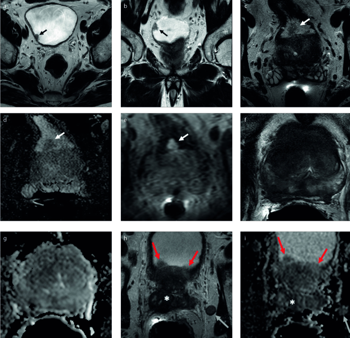
Results: There were 3147 prostate mpMRIs performed during this period and 25 cases (0.8%) of incidental bladder lesions were detected. These patients did not have any presenting symptoms such as gross or microscopic hematuria to prompt bladder lesion workup. The largest diameter of incidentally discovered bladder lesions ranged from 0.4 cm to 1.7 cm. Of the 25 cases of incidental bladder lesions, five were suspected to be due to prostate cancer invasion into the bladder. Only two of these five patients underwent biopsy, which confirmed prostate adenocarcinoma in both cases. Of the 20 patients without suspected prostate cancer invasion of the bladder, four had no suspicious lesions on cystoscopy to warrant a biopsy. The remaining 16 patients had bladder lesions seen on cystoscopy and underwent a biopsy and/or TURBT. Three of these patients had benign features on pathology (urachal remnant, amyloidosis and inflammation) and the remaining 13 had stage Ta urothelial carcinoma. Seven of these patients had low-grade Ta tumors and six had high-grade Ta tumors. All patients were treated with standard management of TURBT with or without intravesical BCG. There have been no reported cases of recurrence or progression in any of the patients in our cohort at the median follow-up of 26 months (interquartile range,19-40 months).
Conclusion: mpMRI of the prostate may yield incidental findings, such as small bladder tumors. Awareness of the possibility of incidental bladder lesions is important as 65% of lesions reported in the bladder, not attributable to extension of prostate cancer, proved to be bladder cancer. This may allow for early intervention for asymptomatic patients with undetected bladder cancer prior to disease progression.
Conflict of interest statement
The authors declared no conflicts of interest.
Figures
References
Publication types
MeSH terms
LinkOut - more resources
Full Text Sources
Other Literature Sources
Medical
