

Long-Term Engraftment (16 Years) of Myoblasts in a Human Infarcted Heart
- PMID: 30211981
- PMCID: PMC6186271
- DOI: 10.1002/sctm.18-0017
Long-Term Engraftment (16 Years) of Myoblasts in a Human Infarcted Heart
Abstract
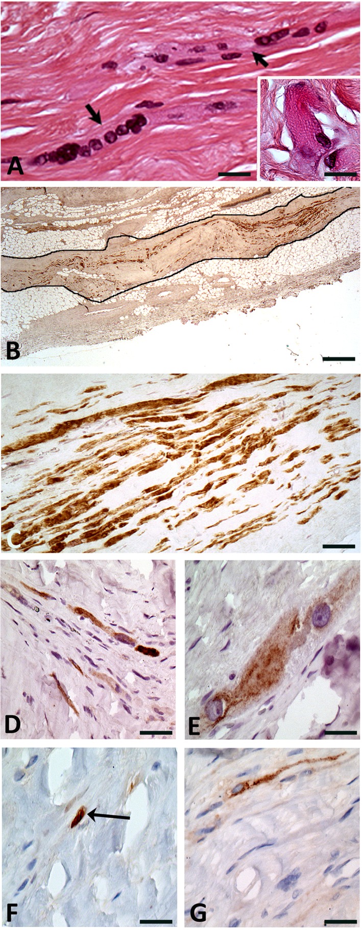
We report the case of a patient who had undergone injections of myoblasts in an infarct area 16 years before being referred for heart transplantation. The pathological examination of the explanted heart found persisting myotubes embedded in fibrosis. This finding supports the ability of myoblasts to survive in harsh environments, which can make them appealing candidates for transplantation in diseases requiring supply of new myogenic cells. Stem Cells Translational Medicine 2018;7:705-708.
Keywords: Clinical cell transplantation; Heart failure; Long-term follow-up; Myoblasts.
© 2018 The Authors. Stem Cells Translational Medicine published by Wiley Periodicals, Inc. on behalf of AlphaMed Press.
Figures
References
-
- Menasché P, Hagège AA, Scorsin M et al. Myoblast transplantation for heart failure. Lancet 2001;357:279–280. - PubMed
-
- Seeger FH, Zeiher AM, Dimmeler S. Cell‐enhancement strategies for the treatment of ischemic heart disease. Nat Clin Pract Cardiovasc Med 2007;4(suppl 1):S110–S113. - PubMed
-
- Skuk D, Goulet M, Roy B et al. First test of a “high‐density injection” protocol for myogenic cell transplantation throughout large volumes of muscles in a Duchenne muscular dystrophy patient: Eighteen months follow‐up. Neuromuscul Disord 2007;17:38–46. - PubMed
MeSH terms
Substances
LinkOut - more resources
Full Text Sources
Other Literature Sources
Medical
