Review
. 2018 Sep 13;14(9):e1007582.
doi: 10.1371/journal.pgen.1007582. eCollection 2018 Sep.

Structure and function of archaeal histones

Affiliations
Review

Structure and function of archaeal histones

Bram Henneman et al. PLoS Genet. .

Abstract

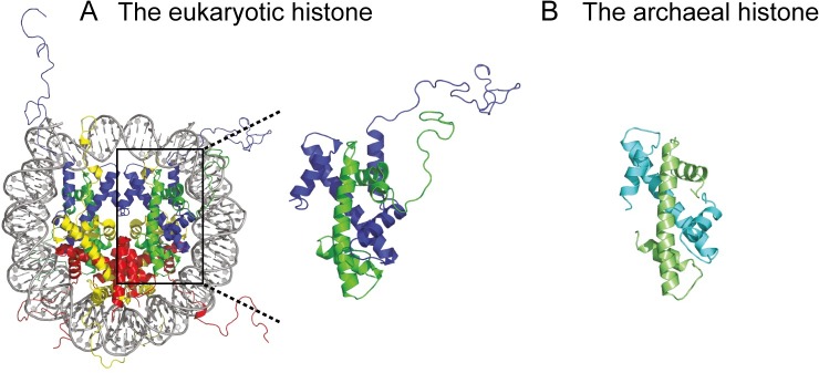
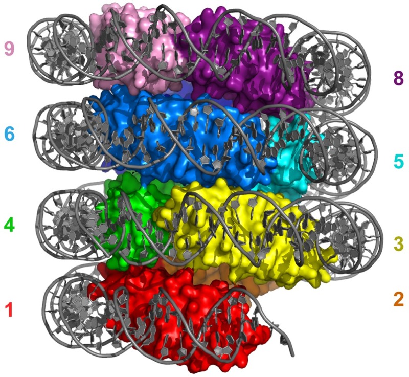
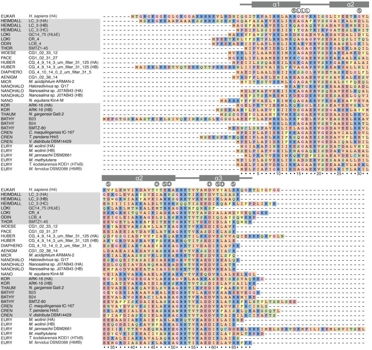
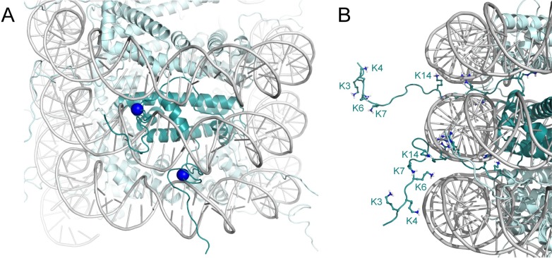
The genomes of all organisms throughout the tree of life are compacted and organized in chromatin by association of chromatin proteins. Eukaryotic genomes encode histones, which are assembled on the genome into octamers, yielding nucleosomes. Post-translational modifications of the histones, which occur mostly on their N-terminal tails, define the functional state of chromatin. Like eukaryotes, most archaeal genomes encode histones, which are believed to be involved in the compaction and organization of their genomes. Instead of discrete multimers, in vivo data suggest assembly of "nucleosomes" of variable size, consisting of multiples of dimers, which are able to induce repression of transcription. Based on these data and a model derived from X-ray crystallography, it was recently proposed that archaeal histones assemble on DNA into "endless" hypernucleosomes. In this review, we discuss the amino acid determinants of hypernucleosome formation and highlight differences with the canonical eukaryotic octamer. We identify archaeal histones differing from the consensus, which are expected to be unable to assemble into hypernucleosomes. Finally, we identify atypical archaeal histones with short N- or C-terminal extensions and C-terminal tails similar to the tails of eukaryotic histones, which are subject to post-translational modification. Based on the expected characteristics of these archaeal histones, we discuss possibilities of involvement of histones in archaeal transcription regulation.


Conflict of interest statement

The authors have declared that no competing interests exist.

Figures

Fig 1
Fig 1. Eukaryotic and archaeal histones.
(A) Eukaryotic nucleosome consisting of DNA wrapped around a core of a (H3-H4)2 tetramer and two H2A-H2B dimers. Yellow, H2A; red, H2B; blue, H3; green, H4. (B) Archaeal histone homodimer of HMfB. HMfB, Histone B from Methanothermus fervidus.
Fig 2
Fig 2. Overview of the hypernucleosome structure.
HMfB dimers stack to form a continuous, central protein core that wraps the DNA in a left-handed superhelix. Nine HMfB dimers are shown, each dimer in surface mode and in rainbow colors. Numbering indicates position of the nine histone dimers; note that dimer 5 and 6 occlude the view of dimer 7. DNA is in gray and shown as cartoon. Image generated using PDB entry 5T5K [64]. HMfB, Histone B from Methanothermus fervidus; PDB, Protein Data Bank.
Fig 3
Fig 3. Alignment of histones from different archaeal species and human histone H4.
Colors indicate the side chain group: R, H, K: blue; D, E: red; A, V, I, L, M: orange; F, Y, W: yellow; S, T, N, Q: green; C: turquoise; G: pink; P: purple. Symbols above the alignment indicate the dimer–dimer interface (D), the loop of the stacking interface (L), and the putative stacking interactions (S) based on HMfB). Secondary structure and numbering of HMfB is used for reference. EUKAR H4, eukaryotic (human) histone H4 NP_724344.1; HEIMDALL LC_3 HA, HB, and HC, Candidatus Heimdallarchaeota OLS22332.1, OLS24873.1, and OLS21974.1, respectively; LOKI GC14_75 HLkE and CR_4, Candidatus Lokiarchaeota KKK41979.1 and OLS16336.1, respectively; ODIN, Candidatus Odinarchaeota OLS18261.1; THOR, Candidatus Thorarchaeota KXH71038.1; WOESE, Candidatus Woesearchaeota OIO61677.1; PACE, Candidatus Pacearchaeota OIO41945.1; HUBER, Candidatus Huberarchaea CG_4_9_14_3_um_filter_31_125 HA and HB, PJB03565.1, and PJB04497.1, respectively; DIAPHERO, Candidatus Diapherotrites PJA17623.1; AENIGM, Candidatus Aenigmarchaeota OIN88081.1; MICR, Candidatus Micrarchaeota Micrarchaeum acidiphilum ARMAN-2 EET90461.1; NANOHALO, Candidatus Nanohaloarchaeota Haloredivivus sp. G17 and Nanosalina sp. J07AB43 HA and HB, EHK01841.1, EGQ42849.1, and EGQ43804.1, respectively; NANO, Nanoarchaeota Nanoarchaeum equitans Kin4-M AAR39197.1; THAUM, Thaumarchaeota Nitrososphaera gargensis Ga9.2 AFU59009.1; BATHY B23, B24, and SMTZ-80, Candidatus Bathyarchaeota KYH36356.1, KYH37304.1, and KON27866.1, respectively; CREN, Crenarchaeota Caldivirga maquilingensis IC-167, Thermofilum pendens Hrk5, and Vulcanisaeta distribute DSM14429, ABW02527.1, ABL77757.1, and ADN51226.1, respectively; EURY, Euryarchaeota Methanobrevibacter wolinii HA and HB, Methanocaldococcus jannaschii DSM2661, Methanococcoides methylutens, Thermococcus kodakarensis KOD1, and Methanothermus fervidus DSM2066, WP_42707783.1, WP_42706862.1, AAB99668.1, KGK98166.1, BAD86478.1, and ADP77985.1, respectively.
Fig 4
Fig 4. Model of a Heimdall LC_3 hypernucleosome with N-terminal tails.
(A) View showing histone tails protruding through the DNA minor grooves. The R17 Cα-atom is shown as a blue sphere to mark the exit point of the tail. (B) Close up of the histone tails with lysine and arginine residues shown as sticks, and N-terminal lysines are labeled. Homodimers of Heimdall LC_3 histone HA are shown in teal; one dimer is highlighted in darker colors. Models are based on the structure of HMfB (PDB entry 5T5K); the tail in the top (bottom) of panel B is modeled in the H3 (H4) tail conformation (PDB entry 1KX5). HA, Histone A; HMfB, Histone B from Methanothermus fervidus; PDB, Protein Data Bank.
Fig 5
Fig 5. Stacking interface between HMfB dimers in the hypernucleosome.
Each dimer i forms stacking interactions with dimer i+2 and i+3, shown here for dimer 6. Residues deemed important are shown in ball-and-sticks and labeled; close stacking of G16 in dimer 6 and 9 is indicated by arrows (Cα shown as spheres); hydrogen-bonds are indicated with dashes. Image generated using PDB entry 5T5K [64]. HMfB; Histone B from Methanothermus fervidus; PDB, Protein Data Bank.

References

    1. Dorman CJ. Genome architecture and global gene regulation in bacteria: making progress towards a unified model? Nat Rev Microbiol. 2013;11(5):349–55. 10.1038/nrmicro3007 . - DOI - PubMed
    1. Dame RT, Tark-Dame M. Bacterial chromatin: converging views at different scales. Curr Opin Cell Biol. 2016;40:60–5. 10.1016/j.ceb.2016.02.015 . - DOI - PubMed
    1. Eme L, Spang A, Lombard J, Stairs CW, Ettema TJG. Archaea and the origin of eukaryotes. Nat Rev Microbiol. 2017;15(12):711–23. 10.1038/nrmicro.2017.133 . - DOI - PubMed
    1. Williams TA, Foster PG, Cox CJ, Embley TM. An archaeal origin of eukaryotes supports only two primary domains of life. Nature. 2013;504(7479):231–6. 10.1038/nature12779 . - DOI - PubMed
    1. Cox CJ, Foster PG, Hirt RP, Harris SR, Embley TM. The archaebacterial origin of eukaryotes. Proc Natl Acad Sci U S A. 2008;105(51):20356–61. 10.1073/pnas.0810647105 ; PubMed Central PMCID: PMCPMC2629343. - DOI - PMC - PubMed

Publication types

MeSH terms