. 2018 Sep 13;14(9):e1007237.
doi: 10.1371/journal.ppat.1007237. eCollection 2018 Sep.

CD4+T cells mediate protection against Zika associated severe disease in a mouse model of infection

Affiliations

CD4+T cells mediate protection against Zika associated severe disease in a mouse model of infection

Mariah Hassert et al. PLoS Pathog. .

Abstract

Zika virus (ZIKV) has gained worldwide attention since it emerged, and a global effort is underway to understand the correlates of protection and develop diagnostics to identify rates of infection. As new therapeutics and vaccine approaches are evaluated in clinical trials, additional effort is focused on identifying the adaptive immune correlates of protection against ZIKV disease. To aid in this endeavor we have begun to dissect the role of CD4+T cells in the protection against neuroinvasive ZIKV disease. We have identified an important role for CD4+T cells in protection, demonstrating that in the absence of CD4+T cells mice have more severe neurological sequela and significant increases in viral titers in the central nervous system (CNS). The transfer of CD4+T cells from ZIKV immune mice protect type I interferon receptor deficient animals from a lethal challenge; showing that the CD4+T cell response is necessary and sufficient for control of ZIKV disease. Using a peptide library spanning the complete ZIKV polyprotein, we identified both ZIKV-encoded CD4+T cell epitopes that initiate immune responses, and ZIKV specific CD4+T cell receptors that recognize these epitopes. Within the ZIKV antigen-specific TCRβ repertoire, we uncovered a high degree of diversity both in response to a single epitope and among different mice responding to a CD4+T cell epitope. Overall this study identifies a novel role for polyfunctional and polyclonal CD4+T cells in providing protection against ZIKV infection and highlights the need for vaccines to develop robust CD4+T cell responses to prevent ZIKV neuroinvasion and limit replication within the CNS.


Conflict of interest statement

The authors have declared that no competing interests exist.

Figures
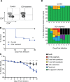
Fig 1
Fig 1. CD4+T cells are necessary for protection from ZIKV challenge.
(A) Confirmation of CD4+T cell depletion. On day –3 and day 0, mice were administered 100 μg of depleting antibody anti-CD4 or isotype control intraperitoneally (n = 9 or 12 mice per group respectively). Blood was harvested at the time of infection and the cells were stained for CD3, CD19, CD4, and CD8. Cells were gated by a lymphocyte gate, CD3+, CD19-, and CD4+ or CD8+. Greater than 99% depletion of CD4+T cells was achieved. (B) Survival of ten- to twelve-week-old Ifnar1-/- mice following CD4+T cell depletion and inoculation with 105 FFU of ZIKV by footpad injection. (n = 9 control, n = 12 depleted). Survival differences were statistically significant (**, p = 0.0063) as determined using a Mantel-Cox test. (C) Weight loss during acute ZIKV infection of ten- to twelve-week-old mice. As a measure of disease, mice were weighed daily for 10 days. On days 7 (*, p = 0.011), 8 (**, p = 0.0039), 9 (**, p = 0.0029), and 10 (***, p = 0.0007) mice from the CD4+ depleted group lost significantly more weight than the control group as determined using an unpaired t test with Welch's correction. (D) Neurological sequela associated with acute ZIKV infection. Mice were evaluated for signs of neurological disease daily and graphed on each day as a percentage of mice displaying that disease indicator. Signs of disease range and in the most severe cases accelerate in the following manner from no apparent disease, limp tail, hind limb weakness, hind limb paralysis, complete paralysis and death. All data is a compellation of 2 independent experiments.
Fig 2
Fig 2. CD4+T cells are important for controlling viral replication in the liver and CNS.
A-F. Viral burden in the peripheral and CNS tissues after CD4+ depletion and ZIKV infection. CD4+ depleted or control mice were infected with 105 FFU ZIKV via footpad injection. On day 4 (n = 4 per group) or day 8 (n = 12–13 per group) post-infection organs were harvested, snap frozen, weighed, and homogenized. Levels of viral RNA were quantified by qPCR in whole blood (A), liver (B), spleen (C), kidney (D), spinal cord (E), and brain (F). Data are shown as Log10 focus-forming unit equivalents (eq.) (as determined by standard curve) per gram or ml of tissue or blood respectively. Data is pooled from 2 independent experiments. Asterisks indicate values that are statistically significant (*, p<0.05, ***, p<0.001) as determined by Mann-Whitney test. (G) Viral RNA was measured by qPCR in whole blood of 10-12-week-old mice over 30 days. Over the time monitored, mice were unable to clear the viral RNA.
Fig 3
Fig 3. CD4+T cell depletion did not alter the CD8+ cellular immune response to ZIKV.
(A) IFNγ production by CD8+T cells in response to stimulation with anti-CD3 or CD8+ epitope E294. On day 8 post-infection, ten- to twelve-week-old depleted or control mice (n = 4 per group) were perfused with 20 ml of PBS. The brains were harvested and lymphocytes were purified as described in the methods section. Lymphocytes were stimulated with CD8+ peptide epitope E294 or anti-CD3 in the presence of brefeldin A, stained and analyzed by flow cytometry. Cells were gated on a lymphocyte gate, CD3+, CD19-, CD4-, CD8+ and were analyzed for functional response by expression of IFNγ. The differences between the control and depleted groups in IFNγ production by CD8+T cells were not statistically significant (p>0.66) as evaluated by Mann-Whitney test. (B) To assess the overall quality of the CD8+T cell response to these stimuli, cells were also analyzed by flow cytometry for IFNγ and TNFα bifunctionality in response to stimulation in the presence of brefeldin A. The differences between the control and depleted groups in CD8+T cell bifunctionality were not statistically significant (p>0.2) as evaluated by Mann-Whitney test. Data is from a single experiment (n = 4).
Fig 4
Fig 4. Identification and functional analysis of ZIKV-specific CD4+T cell epitopes.
(A) ZIKV-specific CD4+T cell epitope identification in the acute phase of infection. C57BL/6J mice (n = 3) were injected IV with 105 FFU of ZIKV. At 10 DPI splenocytes were harvested and stimulated with 10 μg of the indicated peptide in the presence of brefeldin A. Cells were stained for surface markers (CD3, CD19, CD4 and CD8), stained intracellularly for IFNγ and TNFα and analyzed by flow cytometry. Cells were gated using a lymphocyte gate, CD19-, CD4+, CD8- and were functionally analyzed by expression of IFNγ. Data is presented as the percent of CD4+T cells that produced IFNγ in response to stimulation. The dashed line represents the background level of IFNγ production by unstimulated cells. Peptides were classified as hits if the percent of CD4+T cells producing IFNγ was two-fold over background in at least 1 mouse. 18 epitopes were identified, including 4 dominant epitopes. (B) Bifunctionality of response to each epitope. The quality of the response to each epitope during acute infection (n = 3) was evaluated and is represented in the pie charts as the percentage of the total antigen specific cells (IFNγ and/or TNFα positive) which are either positive (gold or grey) or double positive (blue). (C) Functional responses to the immunodominant epitopes are preserved in memory. More than 30 DPI, splenocytes were harvested from infected C57BL/6J mice (n = 3) and were stimulated and stained as described above. Data is presented as the percent of CD4+T cells that produced IFNγ in response to stimulation. The dashed line represents the background level of IFNγ production by unstimulated cells. Data is from a single experiment (n-3).
Fig 5
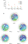
Fig 5. ZIKV-specific CD4+T cell TCR diversity.
(A) TCRβ CDR3 region diversity among CD4+T cells responding to ZIKV-specific epitopes. Wild type C57BL/6J mice (n = 4) were infected with 106 FFU ZIKV. At 10 DPI, splenocytes were harvested and stimulated with the indicated peptide or anti-CD3 in the presence of brefeldin A as described in Fig 4. CD19-, CD8-, CD4+, IFNγ+ cells were sorted separately for each peptide and DNA was extracted for multiplexed amplification of the TCRβ CDR3 region. Unique CDR3 sequences (clonotypes) are enumerated for cells responding to each peptide for each mouse. Each line represents the number of unique TCRβ clonotypes identified from individual peptide or αCD3 stimulated T cells from four mice. (B) Representation of unique TCRβV-J recombinations in cells responding to each stimulation. Circos graphs were generated to display the relative frequency of each specific TCRβV-J join detected in CD4+T cells responding to each epitope. The width of each ribbon is proportional to the number of unique clonotypes that share those specific TCRβV-J segments.
Fig 6
Fig 6. CD4+T cells are sufficient to protect against a lethal ZIKV challenge.
(A) Survival of ten- to twelve-week-old mice following adoptive transfer of CD4+T cells and IV route ZIKV challenge. At 30 days post infection, CD4+T cells were isolated to >97% purity from ZIKV infected or naïve C57BL/6J mice and transferred IV into ten- to twelve-week-old Ifnar1-/- mice (~3x10⁶ /mouse) 1 day prior to IV infection with 105 FFU of ZIKV (n = 9–11 per group). Survival differences were statistically significant between the two groups (p<0.0001) as determined by Mantel-Cox test. (B) Weight loss during IV ZIKV infection of ten- to twelve-week-old mice following adoptive transfer. As a measure of disease, mice were weighed daily for 14 days. No statistically significant differences in weight loss were observed. (C) Neurological sequela associated with IV ZIKV challenge following adoptive transfer. Mice were evaluated for signs of neurological disease daily and graphed on each day as a percentage of mice displaying that disease indicator. Signs of disease range and in the most severe cases accelerate in the following manner from no apparent disease, limp tail, hind limb weakness, hind limb paralysis, complete paralysis and death. Data is pooled from 2 independent experiments.

References

    1. Dick GW, Kitchen SF, Haddow AJ. Zika virus. I. Isolations and serological specificity. Transactions of the Royal Society of Tropical Medicine and Hygiene. 1952;46(5):509–20. . - PubMed
    1. Duffy MR, Chen TH, Hancock WT, Powers AM, Kool JL, Lanciotti RS, et al. Zika virus outbreak on Yap Island, Federated States of Micronesia. The New England journal of medicine. 2009;360(24):2536–43. 10.1056/NEJMoa0805715 . - DOI - PubMed
    1. Chan JF, Choi GK, Yip CC, Cheng VC, Yuen KY. Zika fever and congenital Zika syndrome: An unexpected emerging arboviral disease. The Journal of infection. 2016. 10.1016/j.jinf.2016.02.011 . - DOI - PMC - PubMed
    1. Barton MA, Salvadori MI. Zika virus and microcephaly. CMAJ: Canadian Medical Association journal = journal de l'Association medicale canadienne. 2016;188(7):E118–9. 10.1503/cmaj.160179 ; PubMed Central PMCID: PMC4835294. - DOI - PMC - PubMed
    1. Cauchemez S, Besnard M, Bompard P, Dub T, Guillemette-Artur P, Eyrolle-Guignot D, et al. Association between Zika virus and microcephaly in French Polynesia, 2013–15: a retrospective study. Lancet. 2016. 10.1016/S0140-6736(16)00651-6 . - DOI - PMC - PubMed

Publication types

MeSH terms