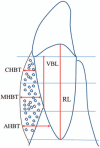
A new modified bone grafting technique for periodontally accelerated osteogenic orthodontics
- PMID: 30212935
- PMCID: PMC6156025
- DOI: 10.1097/MD.0000000000012047
A new modified bone grafting technique for periodontally accelerated osteogenic orthodontics
Abstract
The aim of this study was to introduce an improved surgical technique using a pouch design and tension-free wound closure for periodontally accelerated osteogenic orthodontics (PAOO) in the anterior alveolar region of the mandible.Patients with bone dehiscence and fenestrations on the buccal surfaces of the anterior mandible region underwent the modified PAOO technique (using a pouch design and tension-free closure). Postoperative symptoms were evaluated at 1 and 2 weeks intervals following the procedure. Probing depth (PD), gingival recession depth (GRD), and clinical attachment level (CAL) were assessed at the gingival recession sites at baseline, postoperative 6 and 12 months. Cone-beam computerized tomography (CBCT) was used for quantitative radiographic analyses at baseline, 1 week and 12 months after bone-augmentation procedure.The sample was composed of a total of 12 patients (2 males and 10 females; mean age, 21.9 years) with 72 teeth showing dehiscence/fenestrations and 17 sites presenting with gingival recessions. Clinical evaluations revealed a statistically significant reduction in swelling, pain, and clinical appearance from postoperative week 1 to week 2 (P < .05). Moreover, gingival recession sites exhibited a significant reduction in the GRD and an increase in CAL after surgery with mean root coverage of 69.8% at the end of observation period (P < .01). Both alveolar bone height and width increased after surgery (P < .01) and decreased during the 12-month follow-up (P < .01). However, compared with the baseline records, there was still a significant increase in alveolar bone volume (P < .01).This modified PAOO technique may have advantages in terms of soft and hard tissue augmentation, facilitating extensive bone augmentation and allowing the simultaneous correction of vertical and horizontal defects in the labial aspect of the mandibular anterior area.
Conflict of interest statement
The authors have no conflicts of interest to disclose.
Figures


References
-
- Handelman CS. The anterior alveolus: its importance in limiting orthodontic treatment and its influence on the occurrence of iatrogenic sequelae. Angle Orthod 1996;66:95–109. discussion 109–110. - PubMed
-
- Evangelista K, Vasconcelos Kde F, Bumann A, et al. Dehiscence and fenestration in patients with Class I and Class II Division 1 malocclusion assessed with cone-beam computed tomography. Am J Orthod Dentofacial Orthop 2010;138:133.e131–7.e131. discussion 133–135. - PubMed
-
- Koke U, Sander C, Heinecke A, et al. A possible influence of gingival dimensions on attachment loss and gingival recession following placement of artificial crowns. Int J Periodontics Restorative Dent 2003;23:439–45. - PubMed
-
- Leung CC, Palomo L, Griffith R, et al. Accuracy and reliability of cone-beam computed tomography for measuring alveolar bone height and detecting bony dehiscences and fenestrations. Am J Orthod Dentofacial Orthop 2010;137(4 suppl):S109–19. - PubMed
-
- Thilander BL. Complications of orthodontic treatment. Curr Opin Dent 1992;2:28–37. - PubMed
Publication types
MeSH terms
Substances
LinkOut - more resources
Full Text Sources
Other Literature Sources

