. 2018 Jan-Dec;25(1):1073274818798594.
doi: 10.1177/1073274818798594.

Tricyclic Antidepressants Modulate Stressed Mitochondria in Glioblastoma Multiforme Cells

Affiliations

Tricyclic Antidepressants Modulate Stressed Mitochondria in Glioblastoma Multiforme Cells

Anna M Bielecka-Wajdman et al. Cancer Control. 2018 Jan-Dec.

Abstract

A common feature of solid tumors, including glioblastoma multiforme (GBM), is mitochondrial dysfunction. However, it is reported that the current standard of anti-GBM therapies may potentiate mitochondrial damage and, in effect, support the aggressive character of cancer. As mitochondria are implicated in the modulation of cellular drug sensitivity and chemoresistance mechanisms, activation-stressed mitochondria in GBM cells may represent a new target for anti-GBM therapy that is nontoxic for normal cells.

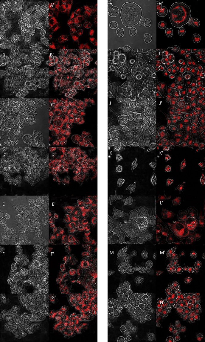
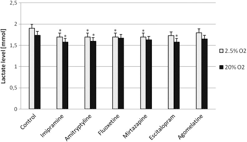
Methods: As mitochondria are possible targets for antidepressant drugs used as adjuvant therapy in patients with GBM, we examined their influence on mitochondrial volume and activity, reactive oxygen species level, extracellular lactate concentration, and p65 NF-κB gene expression in GBM cells.

Results: Our investigation showed, for the first time, that tricyclic antidepressants, imipramine and amitriptyline, partially reverse GBM abnormalities.

Conclusion: In the light of reported studies, the mitochondrial disturbance observed in glioma cells is a dynamic process that can be reversed or silenced. Moreover, imipramine and amitriptyline are attractive cellular metabolic modulators and can potentially be used to restoring a proper function of mitochondria in GBM cells.

Keywords: adjuvants in cancer treatment; antidepressant drugs; metabolic therapy; mitochondrial disturbances in GBM cells.


Conflict of interest statement

Declaration of Conflicting Interests: The author(s) declared no potential conflicts of interest with respect to the research, authorship, and/or publication of this article.

Figures

Figure 1.
Figure 1.
Effect of antidepressant drugs (imipramine (B), amitriptyline (C), fluoxetine (D), mirtazapine (E), escitalopram (F), agomelatine (G) (10 µmol/L)) on mitochondrial localization/morphology of glioma T98G cells cultured in 2.5% oxygen conditions; A, control glioma cells not exposed to antidepressant drugs. Cells were observed using the fluorescent microscope (FV10i, Olympus). Analyses were conducted using the Imaris program (8.2.1) after the incubation of cells with MitoTracker (ThermoFisher Scientific) (denoted by ’). The left panel presents pictures in a contrast-phase microscope. Magnification ×40. Effect of antidepressant drugs (imipramine (I), amitriptyline (J), Fluoxetine (K), mirtazapine (L), escitalopram (M), agomelatine (N) (10 µmol/L)) on mitochondrial localization/morphology of glioma T98G cells cultured in 20% oxygen conditions (standard laboratory conditions); H, control glioma cells not exposed to antidepressant drugs. Cells were observed using the fluorescent microscope (FV10i, Olympus). Analyses were conducted using the Imaris program (8.2.1) after the incubation of cells with mitotracker (ThermoFisher Scientific) (denoted by ’). The left panel presents pictures in a contrast-phase microscope. Magnification ×40.
Figure 2.
Figure 2.
Effect of antidepressants (10 µmol/L) on mitochondrial volume of glioma T98G cells cultured in 2.5% oxygen and standard laboratory conditions (20% oxygen). Data were analyzed by Imaris program (8.2.1). *P < .05 compared with the control.
Figure 3.
Figure 3.
(A and B) Effect of antidepressants (10 µmol/L) on generation of reactive oxygen species (ROS, ROS negative (−) and ROS positive (+) cells) in glioma T98G cells cultured in 2.5% oxygen and standard laboratory conditions (20% oxygen). Data are presented as means ± standard error of the mean of triplicate samples (P = 3). *P < .05 compared to untreated cells.
Figure 4.
Figure 4.
Effect of antidepressants (10 µmol/L) on extracellular level of lactate released from glioma T98G cells cultured in 2.5% oxygen and standard laboratory conditions (20% oxygen). Data points are means ± standard error of the mean of triplicate samples (n = 3); statistical analysis was performed using Student’s t test by comparing antidepressants group versus control (untreated cells). *P < .05 compared with the control (basal) lactate releasing.
Figure 5.
Figure 5.
Effect of antidepressants (10 µmol/L) on p65 nuclear factor κB (NF-κB) gene expression in glioma T98G cells cultured in 2.5% oxygen and standard laboratory conditions (20% oxygen). Data are represented as means ± standard error of the mean of triplicate samples (P = 3). *P < .05 compared with control (100%).

References

    1. Czarnecka AM, Czarnecki JS, Kukwa W, Capello F, Sicińska A, Kukwa A. Molecular oncology focus—is carcinogenesis a mitochondriopathy? J Biomed Sci. 2010;17:31. - PMC - PubMed
    1. Masui K, Cavenee WK, Mischel PS. Cancer metabolism as a central driving force of glioma pathogenesis. Brain Tumor Pathol. 2016;33(3):161–168. - PMC - PubMed
    1. Cairns RA, Harris IS, Mak T. Regulation of cancer cell metabolism. Nat Rev Cancer. 2011;11(2):85–95. - PubMed
    1. Deighton R, Bihan TL, Martin SF, et al. Interactions among mitochondrial proteins altered in glioblastoma. J neurooncol. 2014;118(2):247–256. - PMC - PubMed
    1. Sarosiek KA, Chonghaile TN, Letai A. Mitochondria: gatekeepers of response to chemotherapy. Trends Cell Biol. 2013;23(12):612–619. - PMC - PubMed

MeSH terms