

Intraosseous intraneural perineurioma derived from the inferior alveolar nerve with an abnormality of chromosome 22 and expression of the BCR-ABL fusion gene: report of a case and review of recent literature
- PMID: 30213264
- PMCID: PMC6137890
- DOI: 10.1186/s12957-018-1481-8
Intraosseous intraneural perineurioma derived from the inferior alveolar nerve with an abnormality of chromosome 22 and expression of the BCR-ABL fusion gene: report of a case and review of recent literature
Abstract
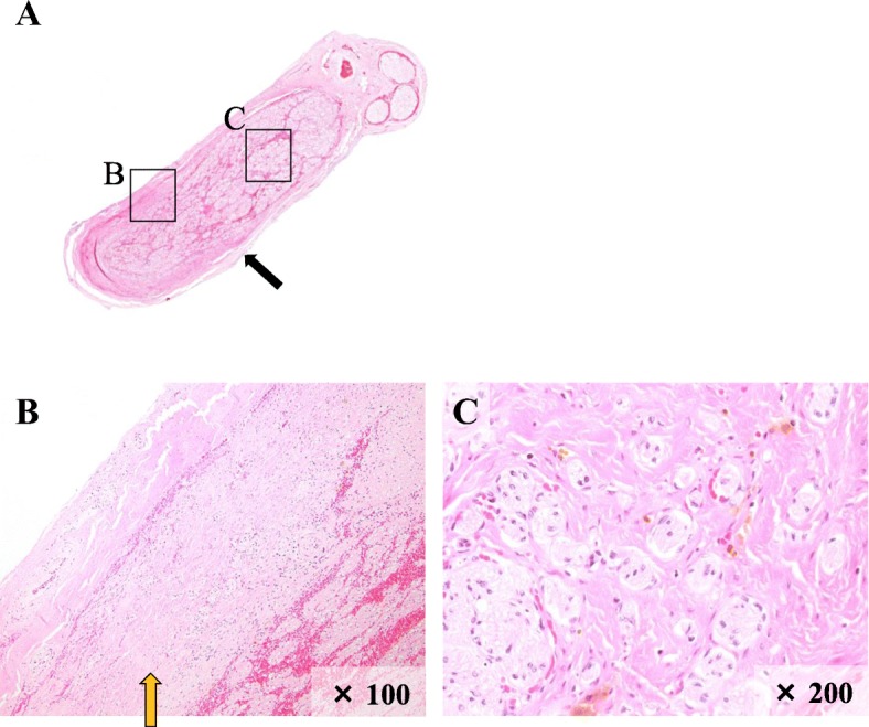
Background: Perineurioma (PN) is a peripheral nerve disease that primarily develops in the limbs and trunk and very rarely occurs in the oral cavity. PN is classified into two types: intraneural perineurioma (INPN) and soft tissue perineurioma (extraneural perineurioma, ENPN). In this article, we report a patient with mandibular body INPN derived from the perineurium of the inferior alveolar nerve.
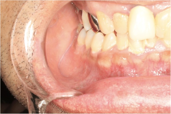
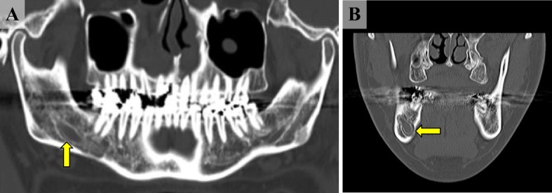
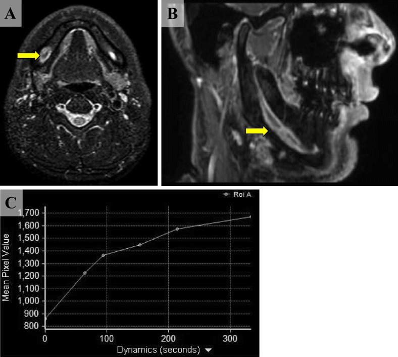
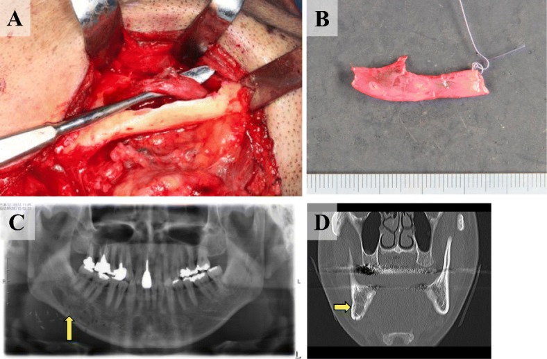
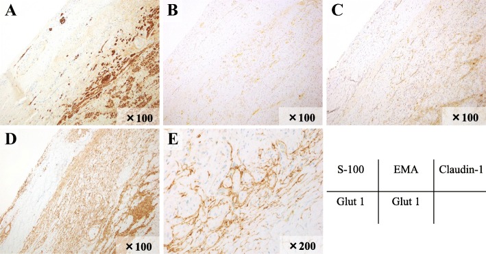
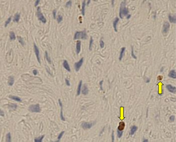
Case presentation: The patient was a 43-year-old male. He consulted our department for a detailed examination of the right mandibular body. A biopsy was performed at another hospital and he was diagnosed with a schwannoma. At his first visit, hypesthesia extending from the right lower lip to the mental region was recognized and enlargement of the right mandibular canal was confirmed with X-ray CT and MRI. Considering the possibility of future tumor growth, we extirpated the tumor under general anesthesia. Cystic tumor was seen continuously in the inferior alveolar nerve. Immunohistologically, the tumor cells were positive for Glut-1, weakly positive for EMA, and weakly positive for Claudin-1, and the histopathological diagnosis was INPN. In addition, absence of the BCR region of chromosome 22 and expression of the BCR-ABL fusion gene were observed by fluorescent in situ hybridization (FISH), and a chromosome 22 abnormality was confirmed. These findings indicated that the disease was a neoplastic lesion.
Conclusion: Expression of the BCR-ABL fusion gene in INPN that develops in the oral cavity is thought to be very rare, and to the best of our knowledge, ours is the first case to be reported in the literature. About three postoperative years have passed, but findings suggestive of recurrence have not been observed.
Keywords: BCR-ABL fusion gene; Chromosome 22 abnormality; ENPN; INPN; Perineurioma.
Conflict of interest statement
Ethics approval and consent to participate
This study was approved by the ethics committee of Gunma University Graduate School of Medicine.
Consent for publication
Informed consent was obtained from the patient.
Competing interests
The authors declare that they have no competing interests.
Publisher’s Note
Springer Nature remains neutral with regard to jurisdictional claims in published maps and institutional affiliations.
Figures

References
-
- Huguet P, De la Torre J, et al. Intraosseous intraneural perineurioma: report of a case with morphological, immunohistochemical and FISH study. Med Oral. 2004;9:64–68. - PubMed
-
- Vencio FE, Cheim PA, Jr, et al. Perineurioma of the mandibular dental nerve: a case report and review of the literature. Oral Surg. 2009;2:103–107. doi: 10.1111/j.1752-248X.2009.01037.x. - DOI
-
- Hata H, Yamazaki Y, et al. Perineurioma of the inferior alveolar nerve with expansion of the mandibular canal. J Oral Maxillofac Surg. 2011;57:419–423.
Publication types
MeSH terms
Substances
LinkOut - more resources
Full Text Sources
Other Literature Sources
Research Materials
Miscellaneous
