Review
. 2018 Oct;39(10):830-847.
doi: 10.1016/j.it.2018.08.006. Epub 2018 Sep 10.

Canonical and Non-Canonical Wnt Signaling in Immune Cells

Affiliations
Review

Canonical and Non-Canonical Wnt Signaling in Immune Cells

Wook-Jin Chae et al. Trends Immunol. 2018 Oct.

Abstract

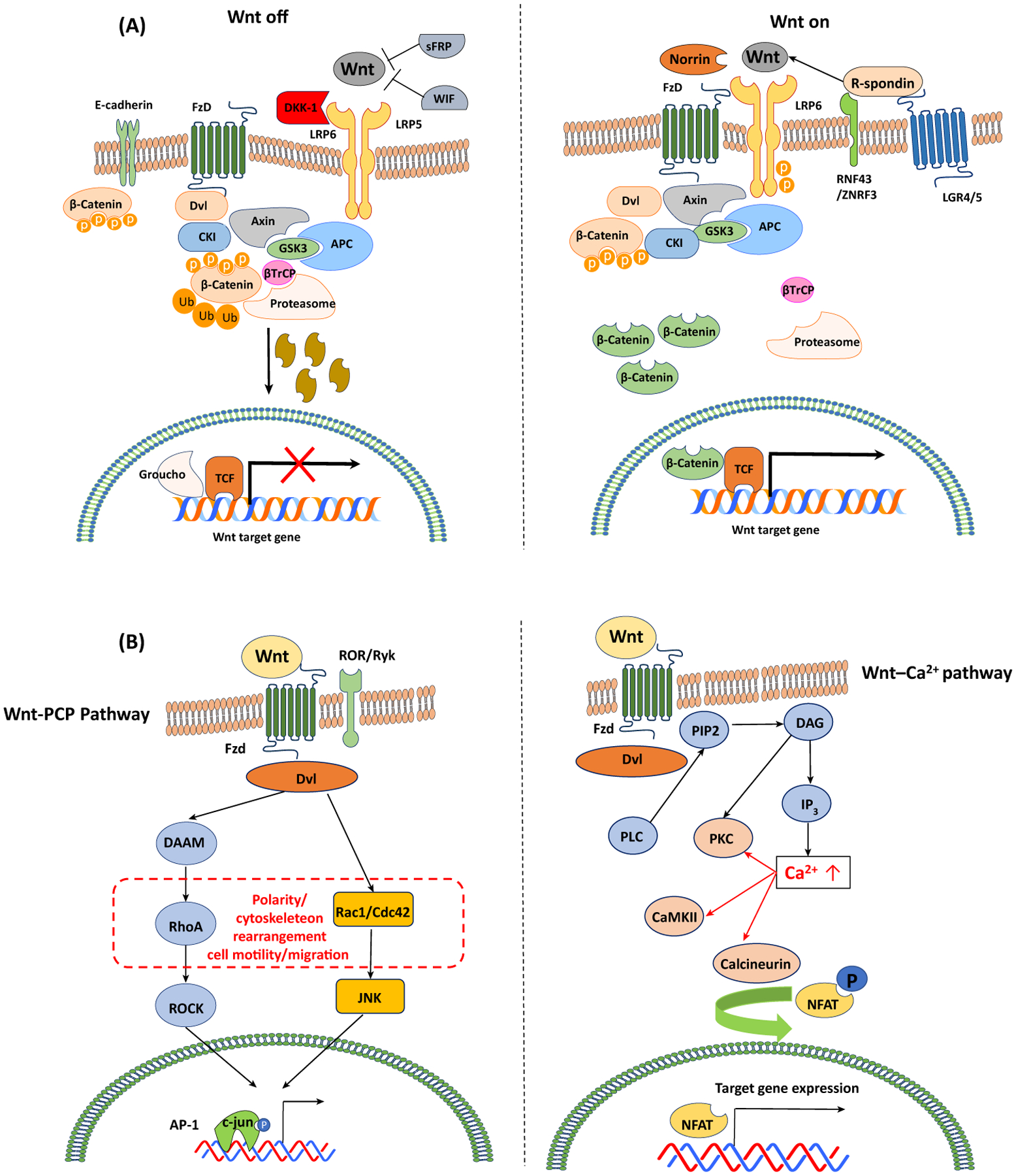
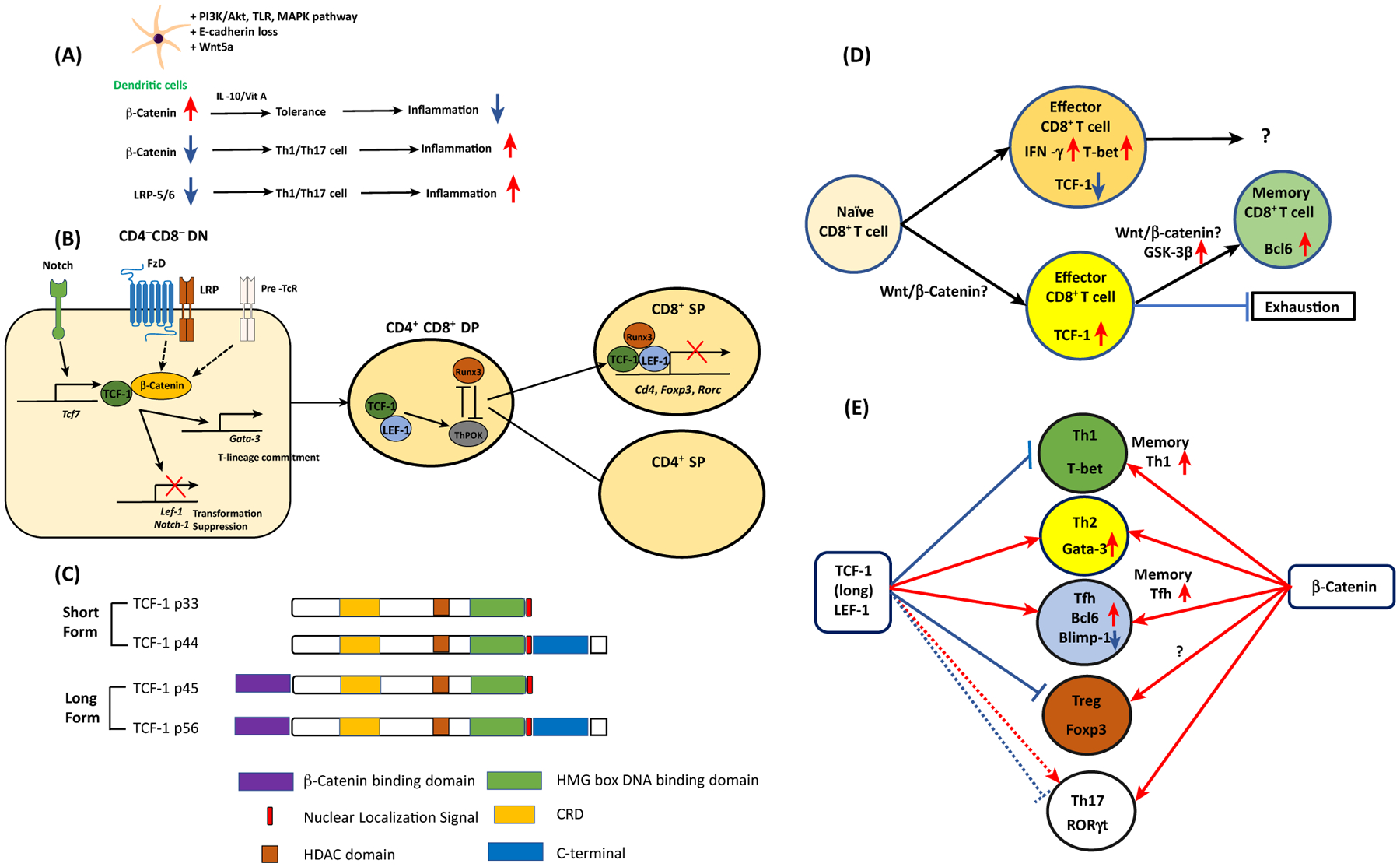
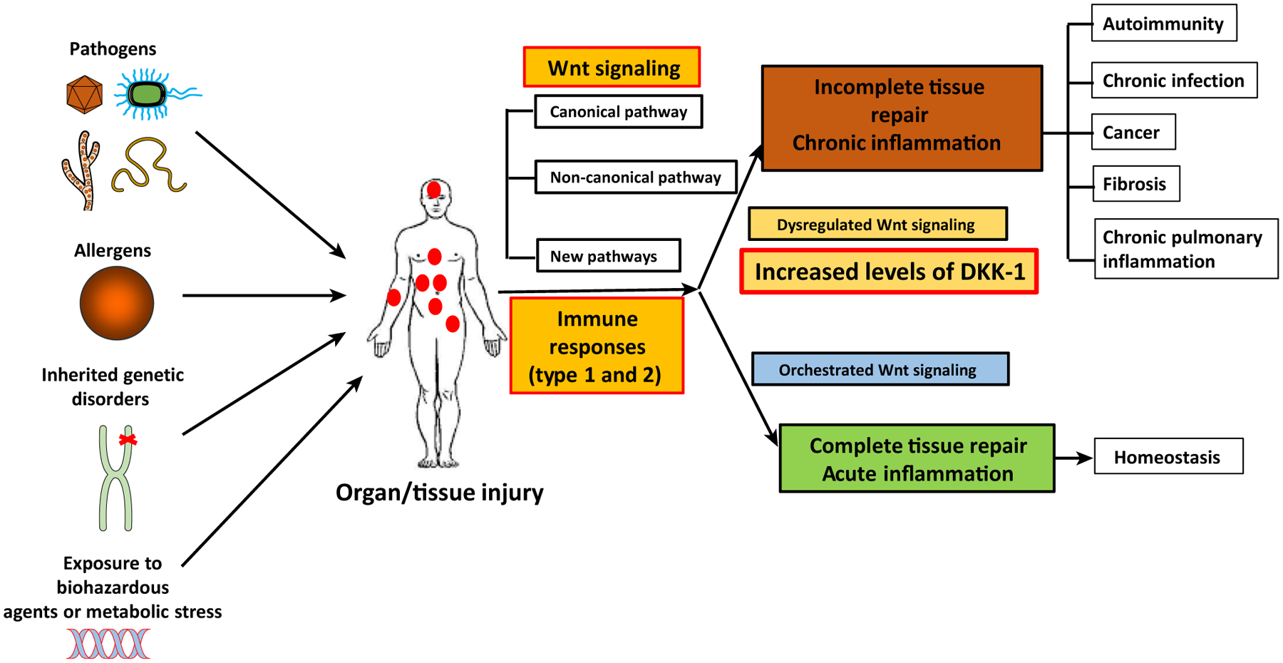
Cell differentiation, proliferation, and death are vital for immune homeostasis. Wnt signaling plays essential roles in processes across species. The roles of Wnt signaling proteins and Wnt ligands have been studied in the past, but the context-dependent mechanisms and functions of these pathways in immune responses remain unclear. Recent findings regarding the role of Wnt ligands and Wnt signaling in immune cells and their immunomodulatory mechanisms suggest that Wnt ligands and signaling are significant in regulating immune responses. We introduce recent key findings and future perspectives on Wnt ligands and their signaling pathways in immune cells as well as the immunological roles and functions of Wnt antagonists.


Figures

Figure 1.
Figure 1.. Canonical and Non-Canonical Wnt Pathways in Mammals (A) Degradation of phosphorylated (P) β-catenin is mediated by the ubiquitin pathway that involves interactions between β-transducin repeat-containing protein (β-TrCP) and the β-catenin destruction complex.
The elimination of cellular β-catenin depletes its pool to translocate to the nucleus; thus, Wnt target gene expression is repressed (Wnt-OFF). Upon Wnt ligand binding to Frz and LRP5/6, the activation of the adaptor protein disheveled (Dvl) and recruitment of Axin complex to the LRP coreceptor are initiated. The decreased proteasomal degradation of phosphorylated β-catenin leads to the accumulation and nuclear translocation of β-catenin, subsequently forming molecular complexes with TCF-1/LEF-1. This interaction eliminates transcriptional repressors (e.g., Groucho), initiating the expression of Wnt target genes (Wnt-ON). Wnt signaling is enhanced by R-spondin binding to its receptor LGR-5 and RNF43. (B) In the Wnt/Ca2+ pathway, Wnt ligands induce the release of intracellular calcium. The Wnt/Ca2+ pathway involves activation of phospholipase C (PLC) and turnover of phospholipid membranes in the endoplasmic reticulum (ER), and the release of intracellular Ca2+. The increased Ca2+ levels result in the activation of Ca2+-dependent signaling molecules such as protein kinase C (PKC), Ca2+/calmodulin-dependent protein kinase (CamKII), and the calcineurin-dependent transcriptional nuclear factor of activated T cells (NFAT). In the planar cell polarity (PCP) pathway, Wnt ligands stimulate activation of the small GTPases Rho and Rac, leading to cytoskeletal rearrangements and cell motility. Ca2+–NFAT signaling plays crucial roles in lymphocyte activation. Signaling crosstalk between the Wnt/Ca2+ pathway and the canonical Wnt pathway to regulate NFAT translocation via GSK-3β has been proposed.
Figure 2.
Figure 2.. Wnt Proteins in T cells and Dendritic Cells (DCs) in Mice and Humans.
(A) β-Catenin accumulation by various signaling pathways and Wnt5a induces tolerogenic DCs to suppress inflammation. When DCs lack β-catenin or Wnt coreceptors LRP5/6, CD4+ T cells are preferentially differentiated into type 1/17 T helper (Th1/Th17) cells to promote inflammation. (B) In the CD4CD8 DN stage, thymocytes receive Notch signals and induce TCF-1. Together with β-catenin, TCF-1 inhibits thymocyte transformation while directing T-lineage commitment via Gata-3 expression. In the CD4CD8 DP stage, TCF-1 interacts with RUNX3 and ThPOK. The interaction between TCF-1/LEF-1 and RUNX3 directs the development of CD8+ T cells. This transcriptional factor complex represses Cd4, Foxp3, and Rorc gene expression. (C) TCF-1 short and long isoforms and their domains. (D) Naïve CD8 T cells are resistant to become CD8 effector T cells if they are TCF-1hi cells, while TCF-1lo cells become active IFN-γ+ T-bet+ effector T cells. TCF-1hi CD8+ effector T cells become more stem-cell-like memory CD8+T cells with elevated expression of Bcl6. Whether the canonical Wnt pathway is involved in this process is controversial. (E) In CD4+ T cells, naïve CD4+ T cells can be differentiated into five different types of helper T cells according to their lineage-specification transcriptional factors. TCF-1 expression favors Th2 and follicular helper T (Tfh) cell formation while it inhibits Th1 differentiation and Treg suppressor function. The role of TCF-1 in Th17 cells is dependent on the microenvironment. In memory CD4+ T cells, TCF-1 enhances memory Th1 and Tfh cell formation. Unlike TCF-1, expression of β-catenin enhances all types of T cell differentiation and function. Abbreviations: DN, double negative; DP, double positive.
Figure 3.
Figure 3.. Wnt Signaling Pathways, Inflammation, and Tissue Repair in Mice and Humans.
A plethora of challenges (e.g., viruses, helminths, fungus, bacteria, allergens, carcinogens, and inherited genetic mutations) cause injuries in various organs and tissues in the body. Upon injury, the host initiates tissue/organ repair by activating Wnt signaling pathways. The canonical Wnt pathway is a central axis of the Wnt immunology. However, recent studies reveal that non-canonical Wnt pathways and non-Wnt pathways that are induced by Wnt ligands are important for type 1 and type 2 core immune responses in tissue injury/repair processes. If tissue repair and immune responses are well coordinated, the host will achieve tissue/organ homeostasis. When these two processes are dysregulated, the host develops multiple chronic inflammatory diseases such as chronic infections, cancer, autoimmune diseases, pulmonary diseases (asthma), and organ fibrosis. Thus, new roles of Wnt ligands and signal transduction pathways are important targets of immunotherapy in many immunological diseases.

References

    1. Nusse R and Clevers H (2017) Wnt/beta-catenin signaling, disease, and emerging therapeutic modalities. Cell 169, 985–999 - PubMed
    1. Karin M and Clevers H (2016) Reparative inflammation takes charge of tissue regeneration. Nature 529, 307–315 - PMC - PubMed
    1. Gieseck RL 3rd et al. (2018) Type 2 immunity in tissue repair and fibrosis. Nat. Rev. Immunol 18, 62–76 - PubMed
    1. Malladi S et al. (2016) Metastatic latency and immune evasion through autocrine inhibition of WNT. Cell 165, 45–60 - PMC - PubMed
    1. Chae WJ et al. (2016) The Wnt antagonist Dickkopf-1 promotes pathological type 2 cell-mediated inflammation. Immunity 44, 246–258 - PMC - PubMed

Publication types