

Geobiology reveals how human kidney stones dissolve in vivo
- PMID: 30213974
- PMCID: PMC6137216
- DOI: 10.1038/s41598-018-31890-9
Geobiology reveals how human kidney stones dissolve in vivo
Abstract
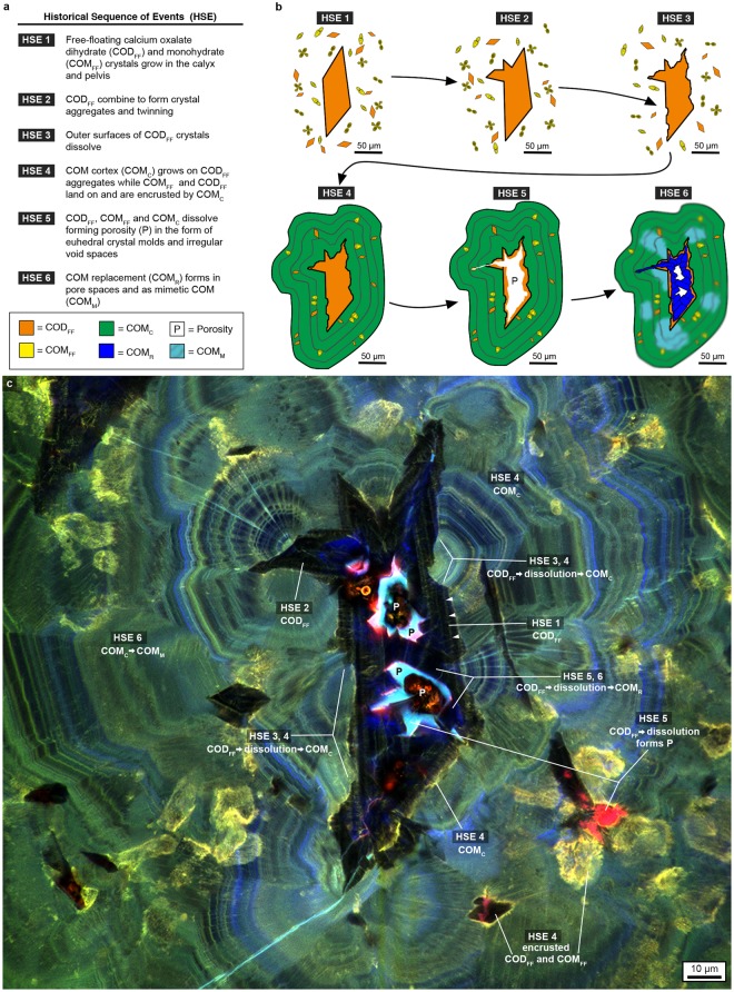
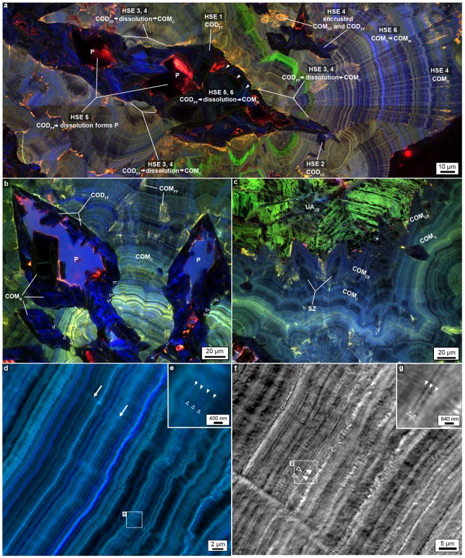
More than 10% of the global human population is now afflicted with kidney stones, which are commonly associated with other significant health problems including diabetes, hypertension and obesity. Nearly 70% of these stones are primarily composed of calcium oxalate, a mineral previously assumed to be effectively insoluble within the kidney. This has limited currently available treatment options to painful passage and/or invasive surgical procedures. We analyze kidney stone thin sections with a combination of optical techniques, which include bright field, polarization, confocal and super-resolution nanometer-scale auto-fluorescence microscopy. Here we demonstrate using interdisciplinary geology and biology (geobiology) approaches that calcium oxalate stones undergo multiple events of dissolution as they crystallize and grow within the kidney. These observations open a fundamentally new paradigm for clinical approaches that include in vivo stone dissolution and identify high-frequency layering of organic matter and minerals as a template for biomineralization in natural and engineered settings.
Conflict of interest statement
The authors declare no competing interests.
Figures
Comment in
-
Re: Geobiology Reveals How Human Kidney Stones Dissolve In Vivo.Eur Urol. 2019 Mar;75(3):532. doi: 10.1016/j.eururo.2018.11.004. Epub 2018 Nov 16. Eur Urol. 2019. PMID: 30449699 No abstract available.
-
Re: Geobiology Reveals how Human Kidney Stones Dissolve In Vivo.J Urol. 2019 Apr;201(4):663-664. doi: 10.1097/01.JU.0000553253.71668.e3. J Urol. 2019. PMID: 30653006 No abstract available.
-
RE: Geobiology reveals how human kidney stones dissolve in vivo (by: Sivaguru et al. 2018).World J Urol. 2019 Nov;37(11):2543. doi: 10.1007/s00345-019-02816-5. Epub 2019 Jun 7. World J Urol. 2019. PMID: 31175461 No abstract available.
References
-
- Fourcroy AF. Mémoire sur le nombre, la nature et les caractères distinctifs des différents matériaux qui forment les calculs, les bézoards et les diverses concrétions des animaux. Ann Museum. 1802;1:93–113.
-
- Moran, M. E. Urolithiasis-A comprehensive history. (Springer, 2014).
-
- Beale, L. S. Illustrations of the Constituents of Urine Urinary Deposits, and Calculi. (John Churchill, 1858).
-
- Ord WM, Shattock SG. On the microscopic structure of urinary calculi of oxalate of lime. Trans Path Soc London. 1895;46:91–132.
-
- Randall A. Analysis of urinary calculi through the use of the polarizing microscope. J Urol. 1942;48:642–649. doi: 10.1016/S0022-5347(17)70755-0. - DOI
Publication types
MeSH terms
Substances
Grants and funding
LinkOut - more resources
Full Text Sources
Other Literature Sources
