

Microwave treatment of the cornea leads to localised disruption of the extracellular matrix
- PMID: 30213993
- PMCID: PMC6137159
- DOI: 10.1038/s41598-018-32110-0
Microwave treatment of the cornea leads to localised disruption of the extracellular matrix
Abstract
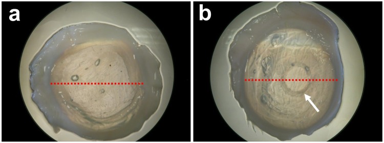
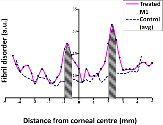
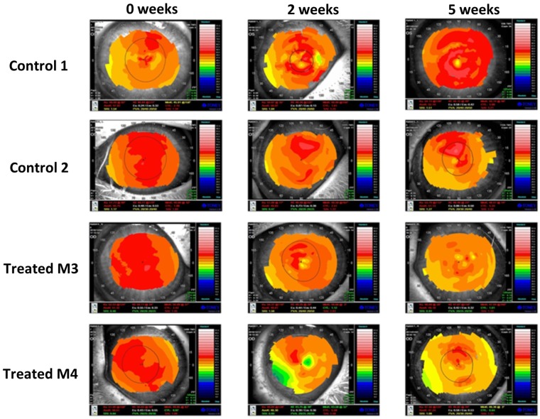
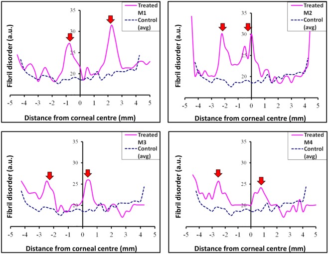
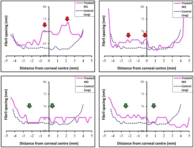
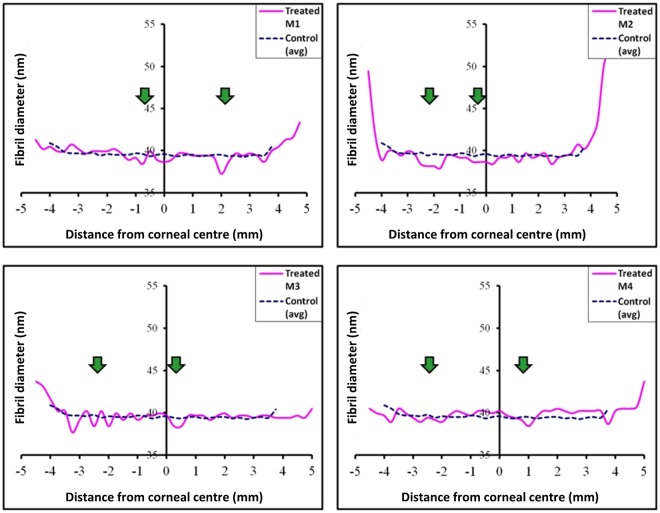
Microwave keratoplasty is a thermo-refractive surgical procedure that can correct myopia (short-sightedness) and pathologic corneal steepening by using microwave energy to cause localised shrinkage around an annulus of the cornea leading to its flattening and vision correction. The effects on the corneal extracellular matrix, however, have not yet been evaluated, thus the current study to assess post-procedure ultrastructural changes in an in-vivo rabbit model. To achieve this a series of small-angle x-ray scattering (SAXS) experiments were carried out across whole transects of treated and untreated rabbit corneas at 0.25 mm intervals, which indicated no significant change in collagen intra-fibrillar parameters (i.e. collagen fibril diameter or axial D-period), whereas inter-fibrillar measures (i.e. fibril spacing and the degree of spatial order) were markedly altered in microwave-treated regions of the cornea. These structural matrix alterations in microwave-treated corneas have predicted implications for corneal biomechanical strength and tissue transparency, and, we contend, potentially render microwave-treated corneas resistant to surgical stabilization using corneal cross-linking procedures currently employed to combat refractive error caused by corneal steepening.
Conflict of interest statement
The authors declare no competing interests.
Figures
References
-
- Lans L. Experimentelle untersuchungen ueber entstehung von astigmatismus durch nicht perforierende corneawunden. Graefes Ophthalmol. 1889;44:117–152.
-
- Flory PJ, Garrett RR. Phase transitions in collagen and gelatin systems. J Am Chem Soc. 1958;80:4836–4845. doi: 10.1021/ja01551a020. - DOI
-
- Deak G, Romhanyi G. The thermal shrinkage process of collagen fibres as revealed by polarization optical analysis of topooptical staining reactions. Acta Morphol Acad Sci Hung. 1967;15:195–208. - PubMed
Publication types
MeSH terms
Substances
Grants and funding
LinkOut - more resources
Full Text Sources
Other Literature Sources
