

Effects of Two-by-Two Combination Therapy with Valproic Acid, Lithium Chloride, and Celecoxib on the Angiogenesis of the Chicken Chorioallantoic Membrane
- PMID: 30214103
- PMCID: PMC6123555
Effects of Two-by-Two Combination Therapy with Valproic Acid, Lithium Chloride, and Celecoxib on the Angiogenesis of the Chicken Chorioallantoic Membrane
Abstract
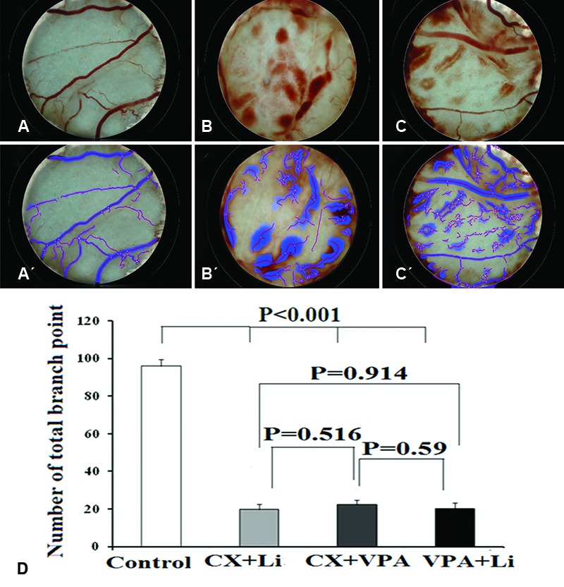
Background: The synergistic effects of valproic acid (VPA), lithium (Li), and celecoxib (CX) have been shown in combination therapy against the proliferation and metastasis of numerous cancers. Angiogenesis plays a critical role in the pathogenesis of tumor growth and metastasis. The aim of the present study was to evaluate the antiangiogenic effects of VPA, lithium chloride (LiCl), and CX, alone or in 2-by-2 combinations, using the chicken chorioallantoic membrane (CAM) assay.
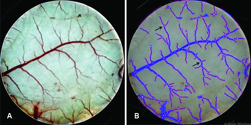
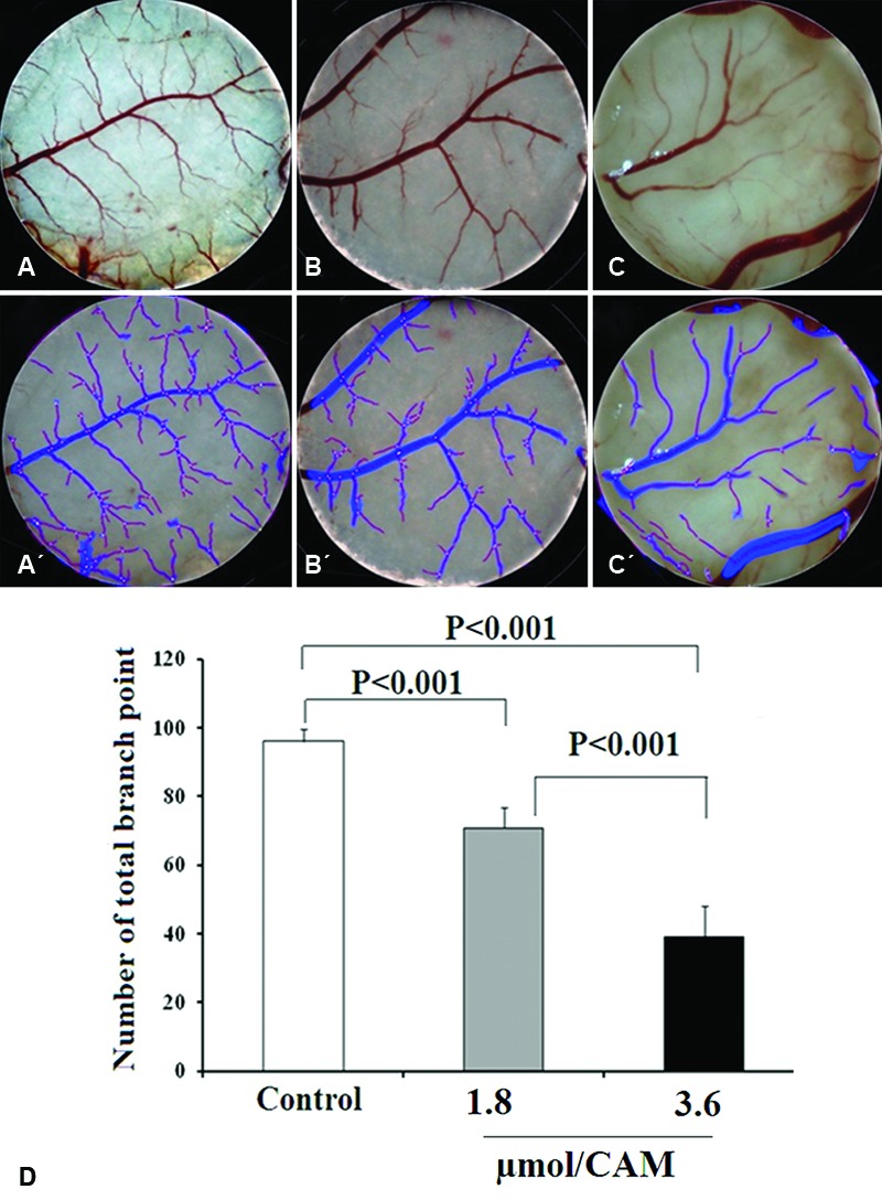
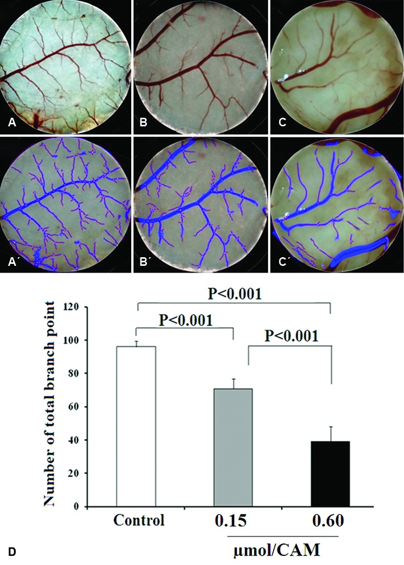
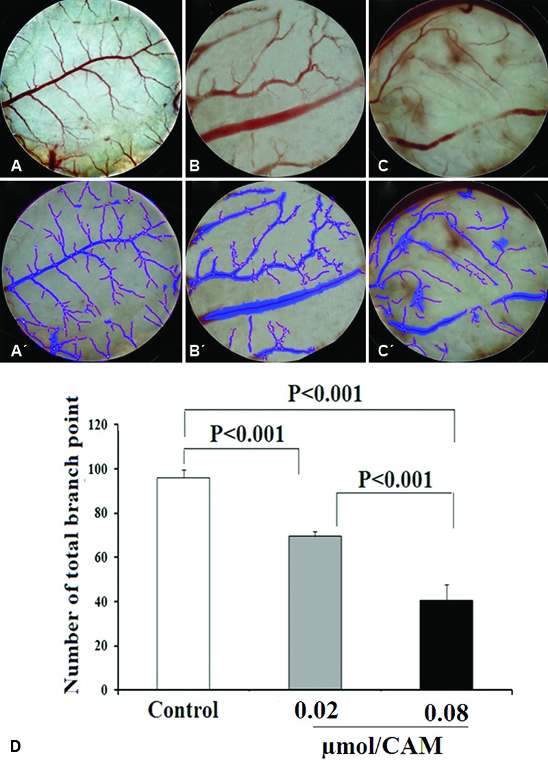
Methods: Fertilized chicken eggs were randomly divided into 10 groups: control, VPA (1.8 and 3.6 µmol/CAM), Li (0.15 and 0.60 µmol/CAM), CX (0.02 and 0.08 µmol/CAM), VPA+Li, VPA+CX, and CX+Li (n=10 per group). A window was made on the eggshells and the CAMs were exposed to a filter disk containing VPA, LiCl, and CX, alone or in 2-by-2 combinations. The control CAMs were treated with distilled water (vehicle). Three days after the treatment, the number of vessel branch points was counted in each CAM. The data were analyzed using SPSS, version 15.One-way ANOVA, followed by the Tukey tests, was used to compare the groups. A P<0.05 was considered a statistically significant difference between the groups.
Results: According to the results, all the tested drugs decreased the number of the vessel branch points in a dose-dependent manner compared to the control group (P<0.001). In addition, combinations of the drugs were more effective in decreasing angiogenesis than the use of each drug alone.
Conclusion: These findings suggest that 2-by-2 combinations of VPA, CX, and LiCl can be considered an effective antiangiogenesis therapeutic modality.
Keywords: Celecoxib; Combined modality therapy; Lithium chloride; Valproic acid; Angiogenesis inhibitors.
Figures
Similar articles
-
Antiangiogenic Activity of n-hexane Insoluble Fraction and Its Tylophorine Component from Ficus septica Leaves in Chicken Chorioallantoic Membrane Induced by bFGF.Asian Pac J Cancer Prev. 2023 Jan 1;24(1):75-80. doi: 10.31557/APJCP.2023.24.1.75. Asian Pac J Cancer Prev. 2023. PMID: 36708554 Free PMC article.
-
A randomized controlled trial comparing lithium plus valproic acid versus lithium plus carbamazepine in young patients with type 1 bipolar disorder: the LICAVAL study.Trials. 2019 Oct 26;20(1):608. doi: 10.1186/s13063-019-3655-2. Trials. 2019. PMID: 31655626 Free PMC article. Clinical Trial.
-
Valproic acid phase shifts the rhythmic expression of Period2::Luciferase.J Biol Rhythms. 2011 Dec;26(6):541-51. doi: 10.1177/0748730411419775. J Biol Rhythms. 2011. PMID: 22215612
-
The Synergistic Effects of Celecoxib and Sodium Valproate on Apoptosis and Invasiveness Behavior of Papillary Thyroid Cancer Cell Line In-vitro.Iran J Pharm Res. 2018 Summer;17(3):1008-1017. Iran J Pharm Res. 2018. PMID: 30127823 Free PMC article.
-
Chick embryo chorioallantoic membrane as a useful tool to study angiogenesis.Int Rev Cell Mol Biol. 2008;270:181-224. doi: 10.1016/S1937-6448(08)01405-6. Int Rev Cell Mol Biol. 2008. PMID: 19081537 Review.
Cited by
-
Predicting Valproate-Induced Liver Injury Using Metabolomic Analysis of Ex Ovo Chick Embryo Allantoic Fluid.Metabolites. 2023 Jun 2;13(6):721. doi: 10.3390/metabo13060721. Metabolites. 2023. PMID: 37367880 Free PMC article.
-
Sodium valproate ameliorates aluminum-induced oxidative stress and apoptosis of PC12 cells.Iran J Basic Med Sci. 2019 Nov;22(11):1353-1358. doi: 10.22038/ijbms.2019.36930.8804. Iran J Basic Med Sci. 2019. PMID: 32128102 Free PMC article.
-
Lithium in Cancer Therapy: Friend or Foe?Cancers (Basel). 2023 Feb 8;15(4):1095. doi: 10.3390/cancers15041095. Cancers (Basel). 2023. PMID: 36831437 Free PMC article. Review.
References
-
- Ribatti D. The discovery of angiogenic growth factors: the contribution of Italian scientists. Vasc Cell. 2014;6:8. doi: 10.1186/2045-824X-6-8. [ PMC Free Article] - DOI - PMC - PubMed
-
- Yadav L, Puri N, Rastogi V, Satpute P, Sharma V. Tumour Angiogenesis and Angiogenic Inhibitors: A Review. J Clin Diagn Res. 2015;9:XE01–XE5. doi: 10.7860/JCDR/2015/12016.6135. [ PMC Free Article] - DOI - PMC - PubMed
-
- Craig MP, Sumanas S. ETS transcription factors in embryonic vascular development. Angiogenesis. 2016;19:275–85. doi: 10.1007/s10456-016-9511-z. [ PMC Free Article] - DOI - PMC - PubMed
-
- Gonzalez AC, Costa TF, Andrade ZA, Medrado AR. Wound healing - A literature review. An Bras Dermatol. 2016;91:614–20. doi: 10.1590/abd1806-4841.20164741. [ PMC Free Article] - DOI - PMC - PubMed
LinkOut - more resources
Full Text Sources