. 2018 Aug 29:10:503-514.
doi: 10.2147/IJWH.S163961. eCollection 2018.

Diet-induced vitamin D deficiency triggers inflammation and DNA damage profile in murine myometrium

Affiliations

Diet-induced vitamin D deficiency triggers inflammation and DNA damage profile in murine myometrium

Heba Elhusseini et al. Int J Womens Health. .

Abstract

Background: Previously, we reported a significantly higher prevalence of uterine fibroids (UFs) in African American women. This minority group also commonly suffers from vitamin D deficiency. We have demonstrated that 1,25(OH)2D3 attains a fibroid growth inhibitory impact through its ability to block the G1/S (gap 1/synthesis) phase of the cell cycle. Vitamin D is involved in DNA damage as well as in immune response regulation, anti-inflammation, autoimmunity and cancer. Since most of the prior data on vitamin D and UF were generated in vitro via established cell lines, it was necessary to verify and validate this observation in vivo using a diet-induced vitamin D-deficient mouse model.

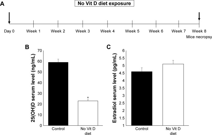
Materials and methods: Our model of vitamin D lacking function was established using 8-week exposure of C57/BL6 mice to vitamin D-deficient diet provides evidence of different functions accomplished by vitamin D in the regulation of myometrium homeostasis disrupted in the context of uterine fibroid.

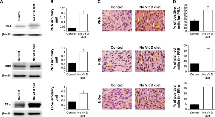
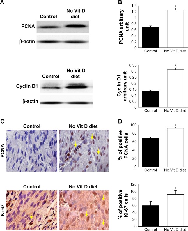
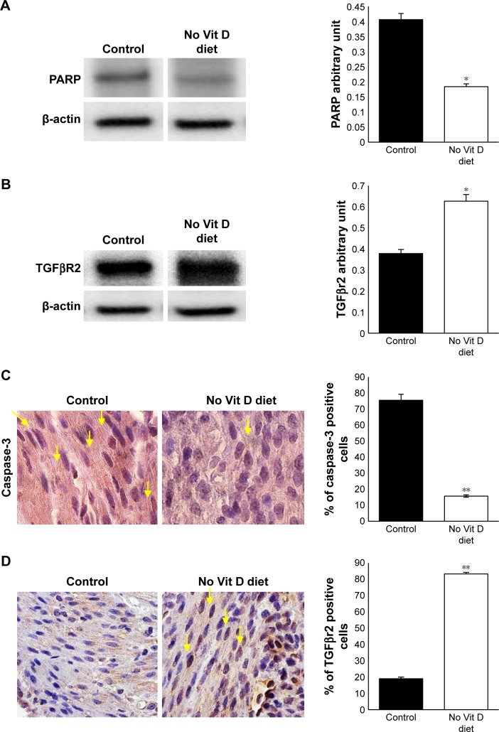
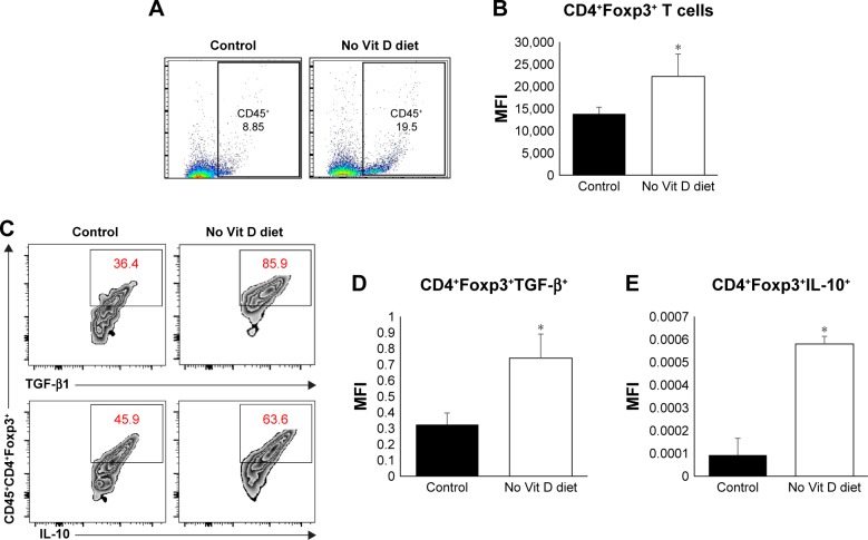
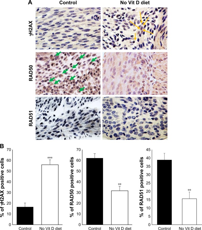
Results: We found that vitamin D deficiency was associated with increased expression of sex steroid receptors in murine myometrium, increased expression of proliferation related genes, the promotion of fibrosis and enhanced inflammation, and promoted immunosuppression through Tregs expansion in murine myometrium. We also showed that a vitamin D deficient diet enhanced DNA damage in murine myometrium.

Conclusion: Our model mimics the effects in humans that a lack of vitamin D has and propels the study of in vivo interaction between inflammation, genomic instability and cell proliferation in the myometrium.

Keywords: DNA damage; diet; inflammation; uterine fibroids; vitamin D.


Conflict of interest statement

Disclosure The authors report no conflicts of interest in this work.

Figures

Figure 1
Figure 1
In vivo levels of vitamin D and estradiol. Notes: (A) Study design of mice exposition to deficient vitamin D diet for a time period of 8 weeks. Mice serum level of vitamin D (B) and estradiol D3 (C) from vitamin D deficiency and control groups (n = 10) were measured using RIA (*P ≤ 0.05). Abbreviation: Vit, vitamin.
Figure 2
Figure 2
Effects of vitamin D deficiency on sex steroid receptors. Notes: (A) Western blot analyses of myometrial cell lysates used to prepare proteins screened with antibodies against PRA, PRB, ER-α and β-actin. (B) Protein expression was quantified by densitometry based on the matched actin expression. (C) Immunohistochemistry staining of embedded fixed myometrium isolated from both groups of mice was prepared as described in the “Materials and methods” section and probed specific mouse anti-PRA, anti-PRB and anti-ER-α. (D) The percentage of positive cells stained compared to that of the controls exposed to regular diet with vitamin D is represented in graph bars. Yellow arrows point to the positive nuclei (n = 10, *P ≤ 0.05, **P ≤ 0.005, ***P ≤ 0.0005). Abbreviations: PRA, progesterone receptor A; PRB, progesterone receptor B; ER-α, ER-alpha; Vit, vitamin.
Figure 3
Figure 3
Antiproliferative effects of vitamin D. Notes: (A) Western blot analyses with mouse anti-PCNA, anti-cyclin D1, marker of cell cycle and anti-β-actin. (B) Protein densitometry analysis was done as described in the Materials and methods section. (C) Immunohistochemical analysis of myometrial tissues of vitamin D deficiency and control groups stained by anti-PCNA and anti-Ki-67. (D) The number of positive stained cells (brown nuclei). Black arrows point to the positive nuclei. All images were captured at 63× magnification, scale bar 200 µm) (*P ≤ 0.05). Abbreviations: PCNA, proliferating cell nuclear antigen; Vit, vitamin.
Figure 4
Figure 4
Vitamin D deficiency inhibits apoptosis and enhances TGFβR2 in myometrium. Notes: (A) Western blot of PARP using mouse anti-PARP antibody. (B) TGFβR2 Western blot using mouse anti-TGFβR2 antibody supported by quantification analysis by densitometry. (C) Immunohistochemical analysis for apoptosis marker caspase-3 using mouse anti-caspase-3. (D) Immunohistochemical analysis for fibrosis in myometrium tissue using mouse anti-TGFβR2. All images were captured at 63× magnification, scale bar 200 µm. The number of stained cells (brown nuclei) was counted in five random high power fields, and the average number of cells is presented in bar graph. Yellow arrows point to the stained nuclei (n = 10, *P ≤ 0.05, **P ≤ 0.005). Abbreviation: Vit, vitamin.
Figure 5
Figure 5
Role of vitamin D in the immune homeostasis of myometrium milieu. Notes: (A) Flow cytometry analysis of myometrium total CD45+ immune cells’ infiltration. (B) MFI analysis after intracellular staining of Foxp3+ Tregs marker gating on CD45+CD4+ T cells. (C) Percentage of positive Tregs for TGF-β1 (upper panels) and IL-10 (lower panels) based on intracellular staining and FACS analysis with MFI of (D) TGF-β1 and (E) IL-10, respectively (n = 10, *P ≤ 0.05). Abbreviations: MFI, mean fluorescence intensity; Tregs, regulatory T cells; Vit, vitamin.
Figure 6
Figure 6
Vitamin D-deficient diet increases DNA damage in mice myometrium. Notes: (A) Immunohistochemical analyses of myometrial tissues of vitamin D deficiency and control groups by DNA damage marker γH2AX and DNA repair genes RAD50 and RAD51. All images were captured at 63× magnification, scale bar 200 µm, yellow arrows indicate YH2AX, green arrows indicate RAD50. (B) The degrees of DNA damage and DNA repair were scored by counting the γH2AX, RAD50 and RAD51 positive nuclei. The percentage of positive cells was calculated based on the number of stained cells, counted in a total of five random high-power fields in the representative myometrium of control and vitamin D3-deficiency samples. The average number of cells is presented (n = 10, **P < 0.005 and ***P < 0.0005). Abbreviation: Vit, vitamin.

References

    1. Catherino WH, Eltoukhi HM, Al-Hendy A. Racial and ethnic differences in the pathogenesis and clinical manifestations of uterine leiomyoma. Semin Reprod Med. 2013;31(5):370–379. - PMC - PubMed
    1. Nesby-O’Dell S, Scanlon KS, Cogswell ME, et al. Hypovitaminosis D prevalence and determinants among African American and white women of reproductive age. Third National Health and Nutrition Examination Survey, 1988–1994. Am J Clin Nutr. 2002;76(1):187–192. - PubMed
    1. Holick MF. Too little vitamin D in premenopausal women: why should we care? Am J Clin Nutr. 2002;76(1):3–4. - PubMed
    1. Sabry M, Halder SK, Allah AS, Roshdy E, Rajaratnam V, Al-Hendy A. Serum vitamin D3 level inversely correlates with uterine fibroid volume in different ethnic groups: a cross-sectional observational study. Int J Womens Health. 2013;5:93–100. - PMC - PubMed
    1. Thota C, Laknaur A, Farmer T, Ladson G, Al-Hendy A, Ismail N. Vitamin D regulates contractile profile in human uterine myometrial cells via NF-kappaB pathway. Am J Obstet Gynecol. 2014;210(4):347.e1–347.e10. - PMC - PubMed

LinkOut - more resources