Case Reports
. 2018 Aug 30:11:193-199.
doi: 10.2147/IMCRJ.S162655. eCollection 2018.

Collagen fiber changes related to keratoconus with secondary corneal amyloidosis

Affiliations
Case Reports

Collagen fiber changes related to keratoconus with secondary corneal amyloidosis

Kaoru Araki-Sasaki et al. Int Med Case Rep J. .

Abstract

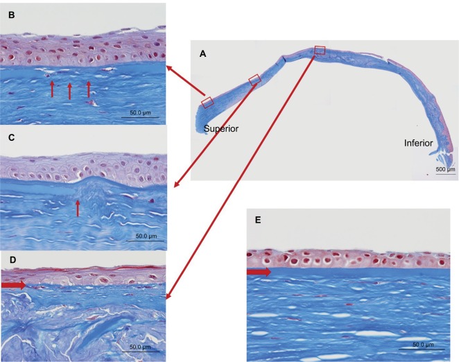
We describe the histological changes in the collagen fibers of a 50-year-old male who presented keratoconus with secondary corneal amyloidosis. Corneal tissue from the patient was obtained following a penetrating keratoplasty and was subjected to histochemical analysis using Masson's trichrome staining, Congo red staining, anti-lactoferrin antibody, and anti-transforming growth factor-beta-induced protein (TGFBIp) antibody. A Congo red-positive region was detected in the anterior half of the stroma in the center and inferior cornea. Although hemotoxylin and eosin staining revealed irregularity in the Congo red-positive region, other parts of the stroma did not show any abnormalities. Positive staining both by anti-TGFBIp and anti-lactoferrin antibodies was observed in the Congo red-positive region. Interestingly, all the layers of the corneal stroma, including the peripheral region, were positively stained by anti-TFGBIp antibody, even in the Congo red-negative area. Masson's trichrome staining also showed irregular staining throughout the corneal stroma, even outside of the Congo red-positive region. Additionally, Bowman's layer, which consists of collagen type IV, was damaged. TGFBIp was strongly expressed and Masson's trichrome staining was reduced throughout the entire keratoconic stroma. The constant qualitative changes in keratoconic collagen fibers, along with the observed abnormality in the Bowman's membrane, might point to the pathogenesis of secondary corneal amyloidosis in keratoconus.

Keywords: TGFBIp; amyloid; cornea; lactoferrin; stroma.


Conflict of interest statement

Disclosure The authors report no conflicts of interest in this work.

Figures

Figure 1
Figure 1
Preoperative slit-lamp photographs. Notes: (A) Slit-lamp photograph of the right eye before surgery. A milky white mass on the tip of the keratoconic cornea was observed. (B) Slit-lamp photograph of the patient’s fellow eye. The keratoconic cornea was observed, and linear opacity under the epithelium was noted instead of a milky white mass.
Figure 2
Figure 2
Congo red staining of the excised cornea. Notes: (A) A positive stained mass was observed in the stroma connected to the inferior peripheral side of the cornea. (B) Inlet showing an enlarged view of the noted area (red arrow). The corneal stroma was normal, and no remarkable pathogenic appearance was observed in the superior peripheral corneal stroma. (C) Inlet showing an enlarged view of the noted area. The Congo red-positive mass resulted in staining in the upper half of the corneal stroma.
Figure 3
Figure 3
Hemotoxylin and eosin staining of the excised cornea. Notes: (A) Irregularity of the collagen fibers was observed in the center of the cornea, in contrast to the regular alignment observed in the superior peripheral cornea. (B) Inlet showing an enlarged view of the peripheral cornea. (C) Inlet showing an enlarged view of the center of the cornea.
Figure 4
Figure 4
Comparison of staining by Congo red, anti-lactoferrin antibody, and anti-TGFBIp antibody. Notes: (A, B) The eosinophilic material was positively stained with Congo red. (C, D) The eosinophilic material was also positively stained with anti-lactoferrin antibody in an area almost overlapping the Congo red-positive region. (E, F). The complete corneal stroma was positively stained by anti-TGFBIp antibody, including the Congo red-negative (arrow) as well as the Congo red-positive area.
Figure 5
Figure 5
Masson’s trichrome staining of the excised cornea. Notes: (A) The excised cornea showed irregular collagen staining (collagen is stained blue). (B) An enlarged view of the superior peripheral cornea. (C) Higher magnification image of the central cornea. The staining was not uniform throughout the cornea and was negative at certain places within the peripheral cornea (B, arrows) as well as in the central cornea (C, arrows).
Figure 6
Figure 6
Masson’s trichrome staining of the Bowman’s layer. Notes: (A) Staining of the excised cornea. (B–D) An enlarged view of each respective inlet. These inlets provide examples of (B) thinning (arrows), (C) cuts (arrow), and (D) complete disappearance (arrow). (E) An image of a normal corneal region for comparison. An arrow indicates Bowman’s layer.

References

    1. Davidson AE, Hayes S, Jardcastle AJ, Tuft SJ. The pathogenesis of keratoconus. Eye. 2014;28(2):189–195. - PMC - PubMed
    1. Galvis V, Sherwin t, Tello A, Merayo J, Barrera R, Acera A. Keratoconus: an inflammatory disorder? Eye. 2015;29(7):843–859. - PMC - PubMed
    1. Wisse RP, Kuiper JJ, Gans R, Imhof S, Radstake TR, Van der Lelij A. Cytokine expression in keratoconus and its corneal microenvironment: a systematic Review. Ocul Surf. 2015;13(4):272–283. - PubMed
    1. Meek KM, Tuft SJ, Huang Y, et al. Changes in collagen orientation and distribution in keratoconus corneas. Invest Ophthalmol Vis Sci. 2005;46(6):1948–1956. - PubMed
    1. Morishige N, Shin-Gyou-Uchi R, Azumi H, et al. Quantitative analysis of collagen lamellae in the normal and keratoconic human cornea by second harmonic generation imaging microscopy. Invest Ophthalmol Vis Sci. 2014;55(12):8377–8385. - PubMed

Publication types

LinkOut - more resources