Review
. 2018 Aug 30:9:1927.
doi: 10.3389/fimmu.2018.01927. eCollection 2018.

Holding All the CARDs: How MALT1 Controls CARMA/CARD-Dependent Signaling

Affiliations
Review

Holding All the CARDs: How MALT1 Controls CARMA/CARD-Dependent Signaling

Mélanie Juilland et al. Front Immunol. .

Abstract

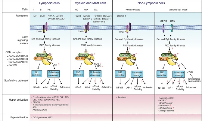
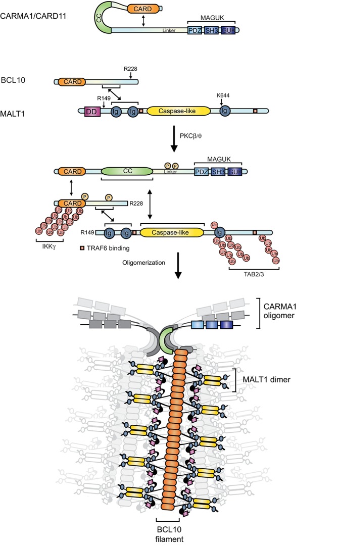
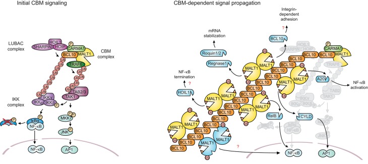
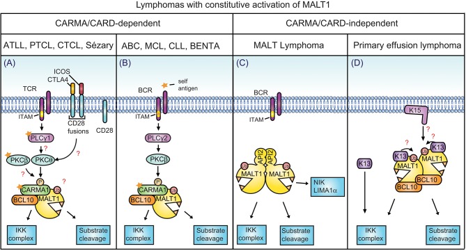
The scaffold proteins CARMA1-3 (encoded by the genes CARD11, -14 and -10) and CARD9 play major roles in signaling downstream of receptors with immunoreceptor tyrosine activation motifs (ITAMs), G-protein coupled receptors (GPCR) and receptor tyrosine kinases (RTK). These receptors trigger the formation of oligomeric CARMA/CARD-BCL10-MALT1 (CBM) complexes via kinases of the PKC family. The CBM in turn regulates gene expression by the activation of NF-κB and AP-1 transcription factors and controls transcript stability. The paracaspase MALT1 is the only CBM component having an enzymatic (proteolytic) activity and has therefore recently gained attention as a potential drug target. Here we review recent advances in the understanding of the molecular function of the protease MALT1 and summarize how MALT1 scaffold and protease function contribute to the transmission of CBM signals. Finally, we will highlight how dysregulation of MALT1 function can cause pathologies such as immunodeficiency, autoimmunity, psoriasis, and cancer.

Keywords: BCR; EGFR; GPCR; RNA stability; TCR; Treg; paracaspase; ubiquitin.


Figures

Figure 1
Figure 1
Signaling via MALT1 downstream of various receptors and CARMA/CARD proteins. CBM complexes composed of MALT1, BCL10 and the indicated CARMA/CARD proteins are formed downstream of receptors containing immunoreceptor tyrosine-based activation motifs (ITAMs), G-protein-coupled receptors (GPCRs) or receptor tyrosine kinases (RTKs). Once formed, the CBM complex activates downstream signaling events via the scaffold and protease function of MALT1. The CBM complex of T and B cells is best characterized, while CBM complexes in other cellular contexts are less well studied. Open questions that remain to be addressed are annotated with a red question mark. Deregulated activation of the pathway results in different diseases, as summarized in the lower panel of the figure. BCR, B cell receptor; DC, dendritic cell; EGFR, epidermal growth factor receptor; FcR, Fc receptor; Mφ, macrophage; MC, myeloid cell; NK, natural killer; OSCAR, osteoclast associated receptor; PKC, protein kinase C; TCR, T cell receptor; TREM-1, triggering expression by myeloid cells 1; RA, rheumatoid arthritis.
Figure 2
Figure 2
Structure and function of the CBM complex. In resting conditions, MALT1 and BCL10 interact constitutively, while CARMA1 is dissociated from the other components of the CBM. PKC-mediated phosphorylation triggers a conformational opening and oligomerization of CARMA1, leading to the recruitment of preformed MALT1- BCL10 complexes. The E3 ligase TRAF6 is further recruited and polyubiquitinates MALT1 and BCL10. This favors the recruitment of the IKK and TAB2/3-TAK1 complexes and the downstream activation of NF-κB and AP-1. N-terminal phosphorylation of BCL10 by IKKβ enhances NF-κB activation, while a C-terminal phosphorylation disassembles MALT1 from BCL10. BCL10 can undergo both, linear polyubiquitination and K63-linked polyubiquitination, which are important for IKKγ/NEMO recruitment. Monoubiquitination activates MALT1 protease function. Once activated, MALT1 cleaves several substrates including BCL10 (after R228) and itself (after R149). The CBM signal is thought to be amplified by formation of filamentous BCL10 structures initiated by CARMA1 nucleation. BCL10 filaments are decorated with MALT1, most likely in its dimeric form. Double-headed arrows indicate interactions. Arrows indicate P, phosphorylations; Ub, ubiquitination.
Figure 3
Figure 3
MALT1 scaffold and protease functions. (A) Antigen receptor triggering leads to the formation of the CBM complex that acts as a recruiting platform for the ubiquitin ligase TRAF6 and the linear ubiquitin chain assembly complex (LUBAC). TRAF6-dependent K63-linked polyubiquitination promotes IKK complex activation by recruiting TAB2/3 together with TAK1 into proximity of the IKK complex. The TAB2/3-TAK1 complex also promotes AP-1 signaling through MKK7 and JNK activation. The CBM complex additionally recruits the LUBAC complex via a CARMA1-HOIP interaction. This is thought to support NF-κB signaling through linear ubiquitination of BCL10, which promotes the physical recruitment of IKKγ/NEMO. (B) Once CBM complex formation has initiated the early signaling events, the signal is most likely amplified and propagated by BCL10 oligomerization and filament formation, and through the protease function of MALT1. Filaments might support the protease function of MALT1 by promoting its dimerization and monoubiquitination. Filaments might also favor or control access of active MALT1 to cellular substrates. The protease activity of MALT1 controls NF-κB activation, AP-1 activation, mRNA stability, and cellular adhesion. This is mediated by the cleavage of the depicted substrates (depicted in blue, also for cleaved BCL10 and autoprocessed MALT1).
Figure 4
Figure 4
CARMA/CARD-dependent and-independent MALT1 activation. (A) In the depicted T cell malignancies, gain-of-function mutations in CARMA1 or its upstream regulators or in-frame gene fusions of the T-cell co-receptor CD28 with ICOS or CTLA-4 are thought to promote MALT1 constitutive activation. (B) In the indicated B-cell lymphomas constitutive MALT1 protease activity can be driven by oncogenic mutations in CARMA1, CD79A or CD79B, which may be relevant in combination with self-antigen recognition. A subset of MCL and CLL are also addicted to this pathway by unknown mechanisms. (C) A large proportion of MALT lymphoma harbors a chromosomal translocation leading to formation of an oncogenic MALT1-API2 fusion protein. (D) In primary effusion lymphoma (PEL), NF-κB is activated through viral latency proteins such as K13 and K15, which activate NF-κB directly or via MALT1-BCL10. (A,B), mutations and self-antigen are depicted with a yellow star. (A,D) Open questions are depicted with red question marks. PTCL, peripheral T-cell lymphoma; ATLL adult T-cell leukemia/lymphoma; ABC, activated B-cell subtype; DLBCL, diffuse large B-cell lymphoma; BENTA, B-cell expansion with NF-κB and T-cell anergy; MCL, mantle cell lymphoma; CLL, chronic lymphocytic/leukemia.

References

    1. Rosebeck S, Rehman AO, Lucas PC, McAllister-Lucas LM. From MALT lymphoma to the CBM signalosome: three decades of discovery. Cell Cycle (2011) 10:2485–96. 10.4161/cc.10.15.16923 - DOI - PMC - PubMed
    1. Getahun A, Cambier JC. Of ITIMs, ITAMs, and ITAMis: revisiting immunoglobulin Fc receptor signaling. Immunol Rev. (2015) 268:66–73. 10.1111/imr.12336 - DOI - PMC - PubMed
    1. Brown GD, Willment JA, Whitehead L. C-type lectins in immunity and homeostasis. Nat Rev Immunol. (2018) 18:374–389. 10.1038/s41577-018-0004-8 - DOI - PubMed
    1. Sun L, Deng L, Ea CK, Xia ZP, Chen ZJ. The TRAF6 ubiquitin ligase and TAK1 kinase mediate IKK activation by BCL10 and MALT1 in T lymphocytes. Mol Cell (2004) 14:289–301. 10.1016/S1097-2765(04)00236-9 - DOI - PubMed
    1. Noels H, van Loo G, Hagens S, Broeckx V, Beyaert R, Marynen P, et al. . A novel TRAF6 binding site in MALT1 defines distinct mechanisms of NF-kappaB activation by API2middle dotMALT1 fusions. J Biol Chem. (2007) 282:10180–9. 10.1074/jbc.M611038200 - DOI - PubMed

Publication types

MeSH terms

Substances