Review
. 2018 Sep 12:12:18.
doi: 10.1186/s13036-018-0108-5. eCollection 2018.

Engineering and monitoring cellular barrier models

Affiliations
Review

Engineering and monitoring cellular barrier models

Jose Yeste et al. J Biol Eng. .

Abstract

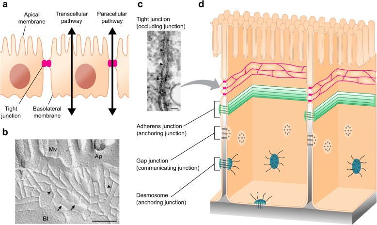
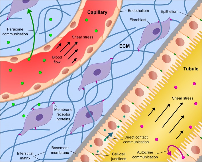
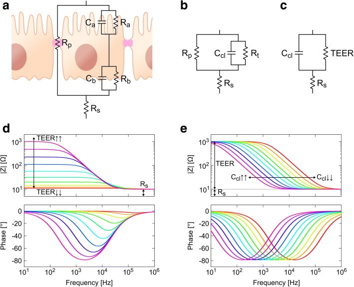
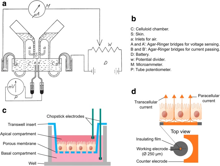
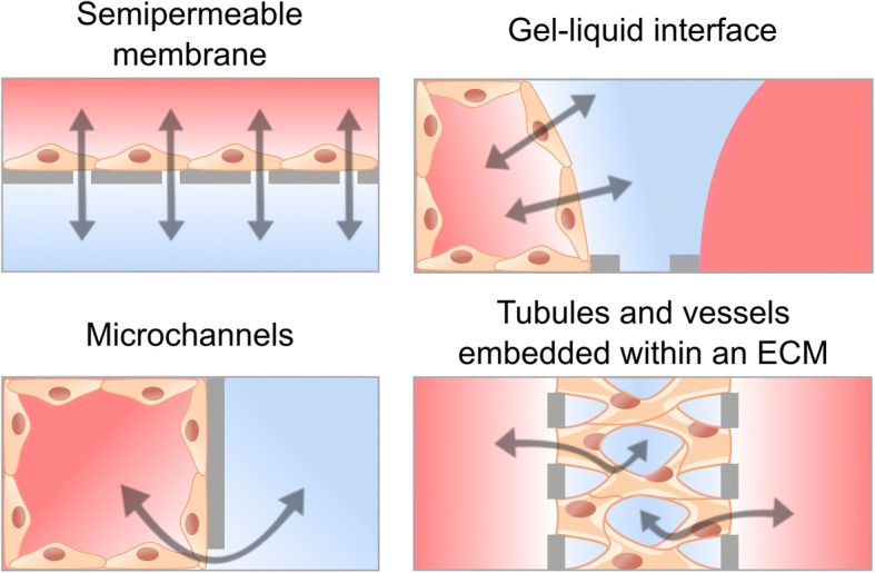
Epithelia and endothelia delineate tissue compartments and control their environments by regulating the passage of ions and solutes. This barrier function is essential for the development and maintenance of multicellular organisms, and its dysfunction is associated with numerous human diseases. Recent advances in biomaterials and microfabrication technologies have evolved in vitro approaches for modelling biological barriers. Current microphysiological systems have become more efficient and reliable in mimicking the cell microenvironment. Additionally, methods for the quantification of barrier permeability have long provided significant insight into their underlying mechanisms. In this review, we outline the current techniques to quantify the barrier function of engineered tissues, and we also give an overview of recent microphysiological systems of biological barriers that emulate the microenvironment and microarchitecture of native tissues.

Keywords: Biological barriers; Cell barrier function; Microphysiological systems; Transepithelial electrical properties.


Conflict of interest statement

Not applicable.Not applicable.The authors declare that they have no competing interests.Springer Nature remains neutral with regard to jurisdictional claims in published maps and institutional affiliations.

Figures

Fig. 1
Fig. 1
Transepithelial transport routes and intercellular junctions. a Paracellular and transcellular pathways across an epithelial layer. b, c Structure and localization of TJ strands including (b) freeze-fracture replica electron microscopic image (scale bar, 200 nm) and (c) ultrathin sectional view (scale bar, 50 nm). Mv, microvilli; Ap, apical membrane; Bl, basolateral membrane. Adapted by permission from Macmillan Publishers Ltd.: Nature Reviews Molecular Cell Biology [1], copyright 2001. d Schematic representation of the intercellular space and the junctional protein complex
Fig. 2
Fig. 2
Physical and biochemical cues in the cell microenvironment. Schematic drawing of an epithelial tubule and an endothelial capillary embedded within an ECM. It includes cell-cell communications (i.e., direct contact, autocrine, and paracrine communications), flow-induced shear stress, ECM involving basement membrane and interstitial matrix components, and cell-ECM interaction through membrane receptor proteins
Fig. 3
Fig. 3
a Equivalent electric circuit for an epithelial cellular layer. b Model with lumped apical and basolateral elements. c Lumped model including only transepithelial electric resistance (TEER) and cell layer capacitance (Ccl). de Representative impedance spectra across a cell layer; data was simulated with Rs equal to 1 kΩ, d TEER values ranging 1–1000 Ω, and e Ccl values ranging 0.1–10 μF. Rp, paracellular resistance; Rs, resistance of the solution; Ra, apical resistance; Rb, basolateral resistance; Ca, apical capacitance; Cb, basolateral capacitance; Rt, transcellular resistance
Fig. 4
Fig. 4
Transepithelial electrical measurement techniques. a Original diagram representation of the Ussing chamber in 1951. Reproduced by permissions of John Wiley and Sons [70]. b Detailed parts of the Ussing chamber. c Schematic representation of chopstick-like electrodes for use with standard Transwell inserts. d Electric Cell Substrate Impedance Sensing system. In this technology, cells are cultured on a surface that contains a small gold electrode (working electrode) and a large counter electrode
Fig. 5
Fig. 5
Compartmentalized approaches for engineering cellular barriers including membrane-based microfluidic device, side-by-side compartments connected through microchannels, gel-liquid interface using phaseguides, and perfusable tubules and microvascular network within an ECM
Fig. 6
Fig. 6
Engineered biological barrier models involving semipermeable membranes or microchannels. a A gut-on-a-chip microfluidic device with spontaneously formation of villi resulting from mechanical cues [107]. TEER profile shows intestinal barrier injury in the presence of pathogenic bacteria (EIEP) or immune cells plus either non-pathogenic bacteria (GFP-EC) or lipopolysaccharide (LPS) endotoxin. b A human kidney proximal tubule-on-a-chip. Immunofluorescence images and bar plots shows increased cell height and increased expression of the tight junction protein ZO-1, aquaporin 1 (AQP1; green), Na/K-ATPase (magenta), and primary cilia in epithelial cells under flow conditions. Adapted from [109] with permission from The Royal Society of Chemistry. c Microfluidic platform for the development of human skin equivalents. Histological and immunofluorescence images demonstrate an improved epidermal morphogenesis and dermoepidermal junction when the tissue is maintained in a dynamic air-liquid interface. Adapted from [110] with permission from Elsevier. d Neonatal BBB model consisted of side-by-side chambers connected through microchannels [114]. Immunofluorescence image shows direct contact communication between endothelial cells (ZO-1; green) and astrocytes (astrocytic marker GFAP; red). e Microfluidic model of the BRB where cells are arranged in parallel compartments and interconnected through a grid of microgrooves [10] – Adapted by permission of The Royal Society of Chemistry. TEER measurement during a calcium switch procedure is performed with two electrodes in the basal side instead of in the apical and basal sides. f Scalable liver-on-a-chip microdevice for long-term maintenance of hepatocyte function in vitro, in which microchannels artificially mimic the fenestrated endothelial cells of the liver [115] (Copyright IOP Publishing. Reproduced with permission. All rights reserved)
Fig. 7
Fig. 7
Engineered biological barrier models involving gel-liquid interface or tubules and vessels embedded within an ECM. a Intestinal epithelium tubule in a microfluidic channel created with a gel interface [118]. As revealed by immunofluorescence, cells form a confluent layer lining the whole channel which results in a perfusable lumen. b Glomerulus-on-a-chip microdevice. Glomerular microtissues are adhered to a gel surface and self-assembled into a continuous barrier of endothelial cells (CD31; red) and podocytes (synaptopodin; green) under flow perfusion. Adapted from [120] with permission from The Royal Society of Chemistry. c Three-dimensional neurovascular microfluidic model that enables heterotypic cell-cell interactions; it comprises human microvascular endothelial cells mimicking cerebral blood vessels, primary rat neurons, and astrocytes. Adapted from [121] with permission from The Royal Society of Chemistry. d Bioprinting method for creating 3D human renal proximal tubules in vitro that are fully embedded within an ECM, including printing of a sacrificial ink, casting of an ECM, evacuation of the ink, and cell seeding [9]. e Three-dimensional bioprinting of thick vascularized tissue consisted of a perfusable vascular network using hUVECs surrounded by hMSCs and fibroblasts [129]. Immunofluorescence image shows osteogenic differentiation (Osteocalcin; violet) of hMSCs in situ after administration of specific growth factors via the vascular network. f A perfusable microvascular network grown in a hydrogel channel. The obtained network exhibits relevant morphological characteristics of in vivo blood vessels. Adapted from [134] with permission from The Royal Society of Chemistry

References

    1. Tsukita S, Furuse M, Itoh M. Multifunctional strands in tight junctions. Nat Rev Mol Cell Biol. 2001;2:285–293. doi: 10.1038/35067088. - DOI - PubMed
    1. Förster C. Tight junctions and the modulation of barrier function in disease. Histochem Cell Biol. 2008;130:55–70. doi: 10.1007/s00418-008-0424-9. - DOI - PMC - PubMed
    1. Zihni C, Mills C, Matter K, Balda MS. Tight junctions: from simple barriers to multifunctional molecular gates. Nat Rev Mol Cell Biol. 2016;17:564–580. doi: 10.1038/nrm.2016.80. - DOI - PubMed
    1. Sawada N. Tight junction-related human diseases. Pathol Int. 2013;63:1–12. doi: 10.1111/pin.12021. - DOI - PMC - PubMed
    1. Ahadian S, Civitarese R, Bannerman D, Mohammadi MH, Lu R, Wang E, et al. Organ-on-a-Chip platforms: A convergence of advanced materials, cells, and microscale technologies. Adv Healthc Mater. 2018;7:1700506. doi: 10.1002/adhm.201700506. - DOI - PubMed