. 2018 Sep 10:10:68.
doi: 10.1186/s13098-018-0370-1. eCollection 2018.

Characterization of spontaneously-developed non-alcoholic fatty liver disease in aged rhesus monkeys

Affiliations

Characterization of spontaneously-developed non-alcoholic fatty liver disease in aged rhesus monkeys

Wen Zheng et al. Diabetol Metab Syndr. .

Abstract

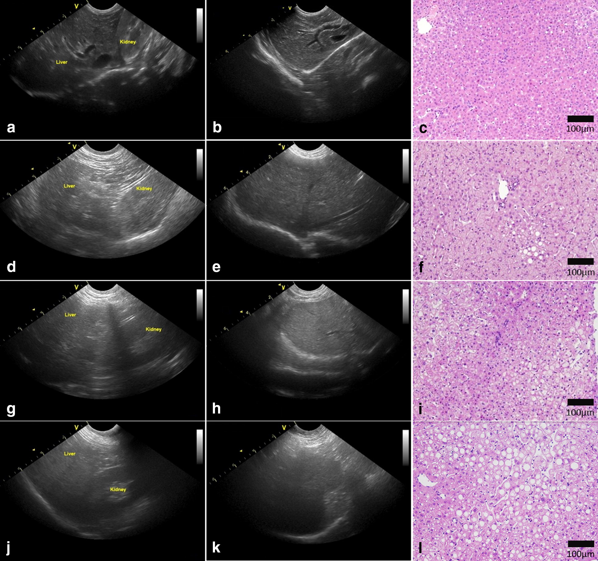
Background: Non-alcoholic fatty liver disease (NAFLD) is a global epidemic afflicting 20-30% in the general population. The animal model of NAFLD available at the present are less clinically relevant. In this study. We aimed to establish a NAFLD model of rhesus monkeys and develop an ultrasonographic steatosis score (USS) system to grade hepatic steatosis in this model.

Methods: We performed hepatic ultrasonography and blood biochemical tests on 86 rhesus monkeys with and without metabolic syndrome (MetS), among which 45 animals were further assessed by histopathological analysis.

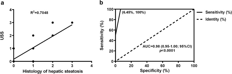
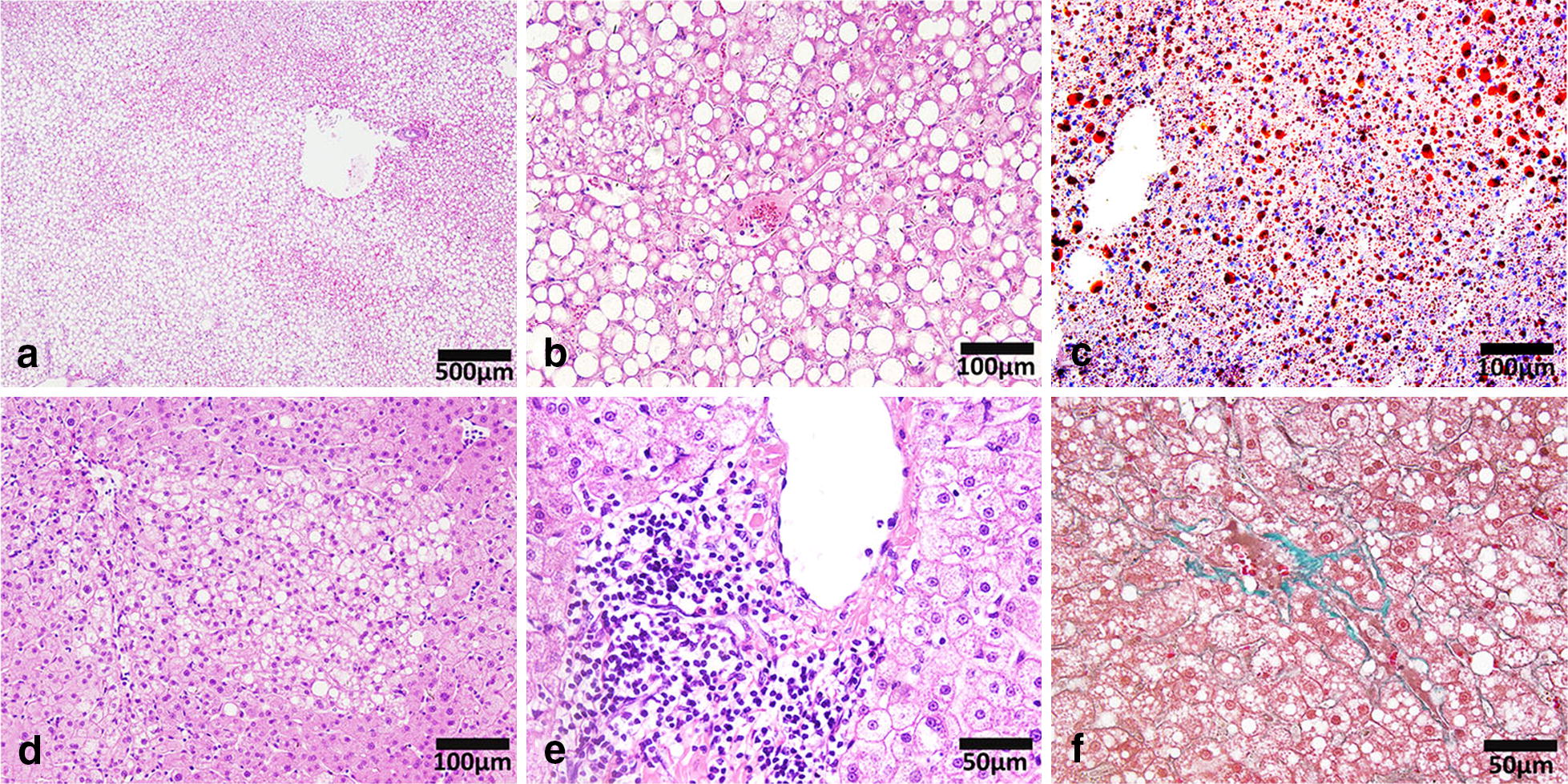
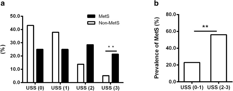
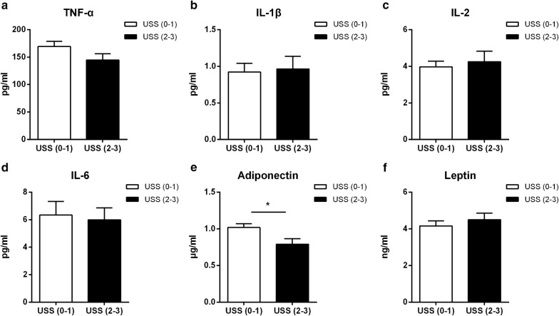
Results: The liver histological features of rhesus monkeys NAFLD were resemble to those of NAFLD patients. There was a close correlation between the histological steatosis grade and the USS (Spearman's coefficient, 0.705, p < 0.001). The USS sensitivity was 87.5% and the specificity was 94.6% when the cut-off was USS2. In addition, the prevalence of MetS was significantly higher in the USS2-3 group. Multiple risk factors of cardiometabolic disease, including obesity, insulin resistance and dyslipidemia were significantly correlated with the USS.

Conclusions: NAFLD was developed spontaneously among aging in rhesus monkeys (with increased prevalence in the MetS monkeys), which provided an ideal model for NAFLD. The newly developed USS system can be used to evaluate fatty liver in the rhesus monkey. The model as well as the noninvasive assessment methodology will provide a powerful tool for mechanistic studies and preclinical test of novel therapies for NAFLD.

Keywords: Metabolic syndrome; Non-alcoholic fatty liver disease; Non-human primates; Ultrasonographic steatosis score.


Figures

Fig. 1
Fig. 1
Representative ultrasonographs and histology. Representative ultrasonographs showing different grades of hepatic steatosis in monkeys. a, b Grade 0, d, e Grade 1, g, h Grade 2, and j, k Grade 3. Representative HE stained sections illustrating a normal liver (c), mild (f), moderate (i), and severe steatosis (l)
Fig. 2
Fig. 2
Correlation between USS and histological steatosis. a Spearman’s correlation between the USS and histological results of steatosis. b Receiver operating characteristic (ROC) curve for the USS to diagnose moderate-to-severe steatosis; cut-off point set at 2 (n = 45; USS ultrasonographic steatosis score)
Fig. 3
Fig. 3
Pathological features of NAFLD in rhesus monkeys. a, b HE staining showing macrovesicular steatosis in monkey liver sections. c Oil Red ‘O’ staining showing the adipose deposition in monkey liver section. d HE staining showing microvesicular steatosis in monkey liver section. e HE staining showing inflammatory cell infiltration in monkey liver sections. f Masson’s trichrome staining showing fibrosis in monkey liver section. (NAFLD non-alcoholic fatty liver disease)
Fig. 4
Fig. 4
Prevalence of hepatic steatosis in MetS monkeys. a Percentages of MetS and non-MetS in normal-to-severe hepatic steatosis groups (non-MetS n = 58; MetS n = 28; *p < 0.05, **p < 0.01). b Prevalence of MetS in moderate-to-severe hepatic steatosis, evaluated by the USS (USS0–1, n = 61; USS2–3, n = 25; *p < 0.05, **p < 0.01; MetS metabolic syndrome, USS ultrasonographic steatosis score)
Fig. 5
Fig. 5
Levels of plasma cytokines and adipokines in different USS groups. a TNF-α. b IL-1β. c IL-2. d IL-6. e Adiponectin. f Leptin. Data are expressed as mean ± SE. USS (0–1), n = 61; USS (2–3), n = 25; *p < 0.05; USS ultrasonographic steatosis score

Similar articles

Cited by

References

    1. Angulo P. Nonalcoholic fatty liver disease. N Engl J Med. 2002;346:1221–1231. doi: 10.1056/NEJMra011775. - DOI - PubMed
    1. Diehl AM, Li ZP, Lin HZ, Yang SQ. Cytokines and the pathogenesis of non-alcoholic steatohepatitis. Gut. 2005;54:303–306. doi: 10.1136/gut.2003.024935. - DOI - PMC - PubMed
    1. Ramesh S, Sanyal AJ. Evaluation and management of non-alcoholic steatohepatitis. J Hepatol. 2005;42(Suppl):S2–S12. doi: 10.1016/j.jhep.2004.11.022. - DOI - PubMed
    1. Choudhury J, Sanyal AJ. Insulin resistance and the pathogenesis of nonalcoholic fatty liver disease. Clin Liver Dis. 2004;8:575–594, ix. doi: 10.1016/j.cld.2004.04.006. - DOI - PubMed
    1. Williams CD, Stengel J, Asike MI, Torres DM, Shaw J, Contreras M, et al. Prevalence of nonalcoholic fatty liver disease and nonalcoholic steatohepatitis among a largely middle-aged population utilizing ultrasound and liver biopsy: a prospective study. Gastroenterology. 2011;140:124–131. doi: 10.1053/j.gastro.2010.09.038. - DOI - PubMed

LinkOut - more resources