. 2018 Sep 12;4(9):eaat7828.
doi: 10.1126/sciadv.aat7828. eCollection 2018 Sep.

Cell fusion potentiates tumor heterogeneity and reveals circulating hybrid cells that correlate with stage and survival

Affiliations

Cell fusion potentiates tumor heterogeneity and reveals circulating hybrid cells that correlate with stage and survival

Charles E Gast et al. Sci Adv. .

Abstract

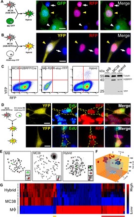
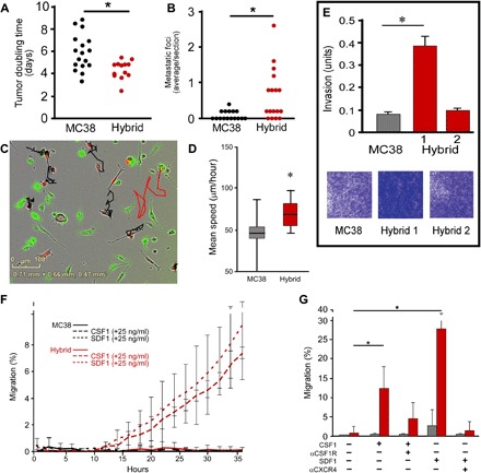
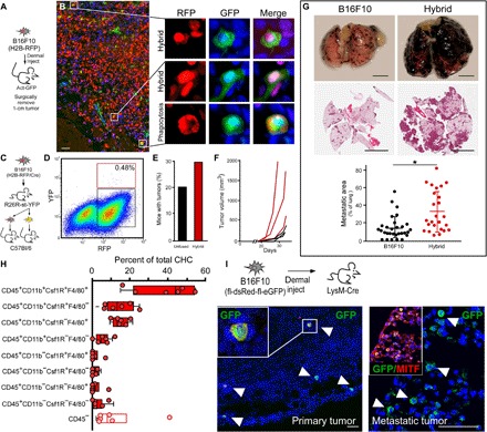
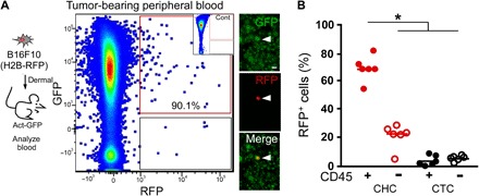
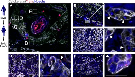
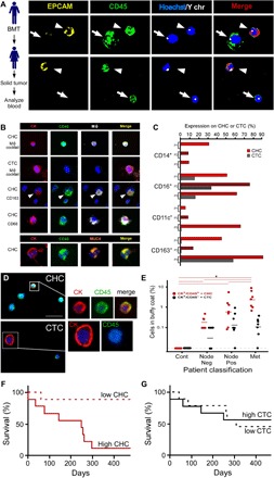
High lethality rates associated with metastatic cancer highlight an urgent medical need for improved understanding of biologic mechanisms driving metastatic spread and identification of biomarkers predicting late-stage progression. Numerous neoplastic cell intrinsic and extrinsic mechanisms fuel tumor progression; however, mechanisms driving heterogeneity of neoplastic cells in solid tumors remain obscure. Increased mutational rates of neoplastic cells in stressed environments are implicated but cannot explain all aspects of tumor heterogeneity. We present evidence that fusion of neoplastic cells with leukocytes (for example, macrophages) contributes to tumor heterogeneity, resulting in cells exhibiting increased metastatic behavior. Fusion hybrids (cells harboring hematopoietic and epithelial properties) are readily detectible in cell culture and tumor-bearing mice. Further, hybrids enumerated in peripheral blood of human cancer patients correlate with disease stage and predict overall survival. This unique population of neoplastic cells provides a novel biomarker for tumor staging, as well as a potential therapeutic target for intervention.


Figures

Fig. 1
Fig. 1. In vitro–derived MФ–cancer cell fusion hybrids.
(A) MC38 (H2B-RFP) cancer cells and GFP-expressing MФs cocultured in a ratio of 1:2 result in hybrid cells with RFP nuclei and GFP-expressing cytoplasm (yellow arrowhead) among unfused cancer cells (white arrow) and MФs (white arrowhead). (B) MC38 (H2B-RFP/Cre) cancer cells cocultured with MФs expressing the Cre reporter, R26R-stop-YFP results in YFP-expressing hybrid cells (yellow arrowhead). (C) YFP-expressing hybrids can be FACS-isolated to purify YFP-expressing hybrid cells confirmed by immunoblot. A representative FACS plot is shown. (D) Cocultured MФs labeled with EdU (green) and MC38 (H2B-RFP/Cre) cancer cells produce YFP-expressing hybrids that initially harbor two nuclei—one from each parent; upon mitotic division, these undergo nuclear fusion, resulting in a single nucleus with EdU-labeled and RFP-expressing DNA. Hybrid cell outlined in yellow. Scale bar, 10 μm. (E) Karyotype and X (red) and Y chromosome (green) fluorescence in situ hybridization (FISH) analyses of parental MФs, unfused MC38 cancer cells, and fusion hybrids. (F) Fusion hybrids (red sphere, n = 45) cluster as a unique population based on their chromosome number and sex chromosomes, relative to MФs (white sphere, n = 27) and MC38s (black sphere, n = 28). (G) Microarray analyses of n = 5 independent hybrid isolates and n = 3 each for MC38 and MФ populations. The yellow bar denotes hybrid gene expression unique from MC38s and MФs. The red bar marks hybrid gene expression that is similar to that in MФs.
Fig. 2
Fig. 2. In vitro–derived fusion hybrid characterization.
(A) Proliferation analysis of MC38 cells and MC38-derived hybrids injected into flanks of immunocompetent syngeneic mice (n = 13 mice, each from two different hybrid isolates). Each data point reflects tumor growth in a single mouse. (B) Analysis of metastatic seeding potential of hybrids and MC38 cells injected into spleens and area analyzed in H&E-stained tissue sections of the liver [n = 15 mice injected with MC38 cells (three different hybrid clones), n = 17 mice injected with hybrids, with each data point reflecting metastatic tumors analyzed in the liver]. (C) Static portrayal of migration tracks from unfused MC38s (black) and a MC38-derived fusion hybrid (red) generated from live-imaged cocultures. Images reflect representative images. (D) The mean speed of hybrids (red bar) relative to MC38s (gray bar) is statistically significant (*P < 1.1 × 10⁻⁹). (E) In vitro invasion assay of MC38 cells and MC38-derived hybrids in Matrigel invasion chambers, stained with crystal violet after 15 hours. Data reflect the average of triplicate samples in biologic replicates. (F) A representative data set evaluating chemotaxis toward CSF1 and SDF1 ligands. Hybrid cell chemotaxis toward CSF1 and SDF1 is statistically significant relative to unfused MC38 cells after 24 hours (P < 0.05). Three independent experiments of triplicates or quadruplicates were conducted for each ligand. Multiple hybrid clones were assessed. (G) Incubation of cells with blocking antibodies to CSF1R and CXCR4 reduces migration of hybrids toward ligands. P < 0.05 and P < 0.01, respectively (hybrid, red bar; MC38, gray bar).
Fig. 3
Fig. 3. B16F10 in vivo–derived fusion hybrids.
(A) B16F10 (H2B-RFP) cells (5 × 10⁴ cells) intradermally injected into GFP-expressing mice (n = 12, two hybrid clones) were harvested at ~1.0 cm at study end point. (B) Fluorescence analyses of tumor sections for RFP (red) and GFP (green) reveal double-positive hybrids and phagocytosed cancer cells with different nuclear morphology. Scale bar, 25 μm. (C) B16F10 (H2B-RFP/Cre) cells injected (5 × 10⁴ cells) into R26R-stop-YFP transgenic mice (n = 8). (D) Representative FACS plot of hybrid and unfused cancer cells from a dissociated tumor, for example, hybrids (red box) and unfused (black box) cancer cells (n = 6 single tumor analyses, n = 2 pooled tumor analyses, n = 13 mice). (E) Three hundred FACS-isolated cells were injected into wild-type secondary recipient mice (n = 19 unfused, n = 19 hybrids) analyzed for tumor growth at 40 days, and (F) 3000 FACS-isolated cells were injected into syngeneic recipient mice (n = 3 MC38 injected mice, black lines; n = 3 hybrid injected mice, red lines) and temporally monitored for growth. (G) B16F10 (H2B-RFP) or MФ–B16F10-derived hybrid cells tail vein–injected into wild-type mice (n = 12 mice). Macroscopic view of lungs and H&E of a tissue section. Quantification of tumor area. (H) Flow analyses of in vivo–derived B16F10 fusion hybrids from a primary tumor. RFP/GFP coexpressing cells analyzed for cell surface MФ identity. All boxes represent hybrid populations. Open box denote hybrids that have lost CD45 expression (n = 6 mice each). (I) B16F10 (fl-dsRed-fl-eGFP) cells intradermally injected into LysM-Cre mice (n = 4) were harvested at ~1 cm. Primary tumor or metastatic lung tumors stained with antibodies to GFP (green) and the tumor protein microphtalmia-associated transcription factor (MITF, red). Scale bar, 25 μm.
Fig. 4
Fig. 4. Murine CTCs.
(A) B16F10 (H2B-RFP) cells (5 × 10⁴ cells) intradermally injected into a syngeneic GFP-expressing recipient mouse. Blood collected at time of tumor resection and analyzed by flow cytometry for GFP and RFP expression. RFP+GFP+ cells were detectible in presorted cell preparations by immunofluorescence. Scale bar, 50 μm. We analyzed GFP-expressing blood by flow cytometry as a negative control for (A) inset. (B) Percentages of fusion hybrids (RFP+/GFP+) and unfused CTCs (RFP+/GFP) expressing the leukocyte antigen CD45 (*P < 0.000002).
Fig. 5
Fig. 5. Cell fusion in human tumors.
Solid tumors from women (n = 7) with previous sex-mismatched bone marrow transplantation (BMT) permits analysis of cell fusion. (A) PDAC tumor section with cytokeratin (gray), the Y chromosome (Y chr, red), and Hoechst (blue) detection revealed areas of cytokeratin-positive cells with Y chr–positive nuclei (white arrowheads). Boxed representative areas are enlarged in (B) to (E). Scale bars, 25 μm.
Fig. 6
Fig. 6. Human CTCs.
(A) Sex-mismatched bone marrow–transplanted (BMT) patient who acquired a solid tumor (PDAC). Peripheral blood was analyzed for the presence of cell fusion. Two panels displaying cell fusion hybrids (arrowheads) that costain for EPCAM (yellow) and CD45 (green) and have a Y chromosome (white dot) in their nuclei (blue). Arrows denote leukocytes. (B) CHCs and CTCs analyzed from n = 4 patients with PDAC. CHCs (CK+/CD45+) also express MФ proteins (cocktail: CD68, CD163, CD66b, and CSF1R), while CTCs (CK+/CD45−ve) do not. CHCs also express the tumor-specific protein MUC4. (C) CHCs and CTCs analyzed by flow cytometry for CD14, CD16, CD11c, and CD163 expression or the cancer-specific protein MUC4 (n = 4 patients). (D) Human pancreatic cancer patient peripheral blood analyzed for cytokeratin+ (red) and CD45+ (green) expression using in situ analyses and digital scanning. (E) CK+/CD45+ and CK+/CD45 cells quantified in patient blood across cancer stages [analysis of variance (ANOVA), *P < 0.023]. (F and G) Kaplan-Meier curve of dichotomized biomarkers based on median value (CHC and CTC) was associated with statistically significant increased risk of death for CHCs (P = 0.0029) but not for CTCs (P = 0.95).

References

    1. Heppner G. H., Tumor heterogeneity. Cancer Res. 44, 2259–2265 (1984). - PubMed
    1. Marusyk A., Polyak K., Tumor heterogeneity: Causes and consequences. Biochim. Biophys. Acta 1805, 105–117 (2010). - PMC - PubMed
    1. Rizvi A. Z., Swain J. R., Davies P. S., Bailey A. S., Decker A. D., Willenbring H., Grompe M., Fleming W. H., Wong M. H., Bone marrow-derived cells fuse with normal and transformed intestinal stem cells. Proc. Natl. Acad. Sci. U.S.A. 103, 6321–6325 (2006). - PMC - PubMed
    1. Davies P. S., Powell A. E., Swain J. R., Wong M. H., Inflammation and proliferation act together to mediate intestinal cell fusion. PLOS ONE 4, e6530 (2009). - PMC - PubMed
    1. Alvarez-Dolado M., Pardal R., Garcia-Verdugo J. M., Fike J. R., Lee H. O., Pfeffer K., Lois C., Morrison S. J., Alvarez-Buylla A., Fusion of bone-marrow-derived cells with Purkinje neurons, cardiomyocytes and hepatocytes. Nature 425, 968–973 (2003). - PubMed

Publication types

MeSH terms

Substances