. 2019 Feb;51(1):29-40.
doi: 10.1007/s10863-018-9772-x. Epub 2018 Sep 13.

Deafferentation-induced alterations in mitral cell dendritic morphology in the adult zebrafish olfactory bulb

Affiliations

Deafferentation-induced alterations in mitral cell dendritic morphology in the adult zebrafish olfactory bulb

Joanna M Pozzuto et al. J Bioenerg Biomembr. 2019 Feb.

Abstract

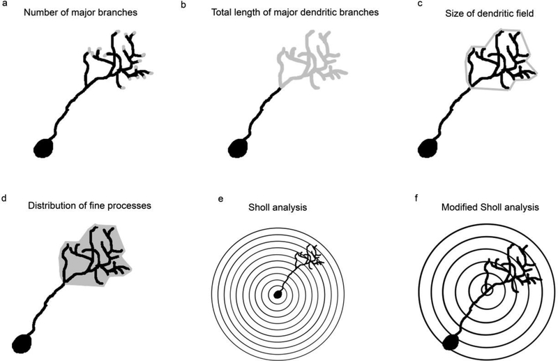
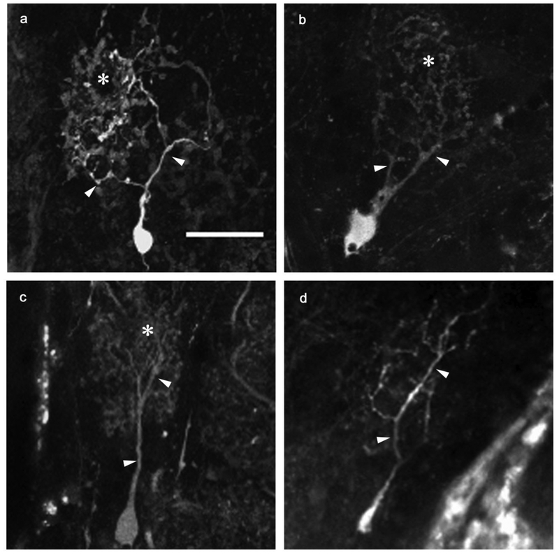
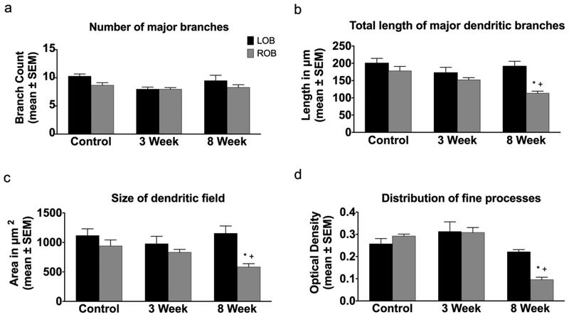
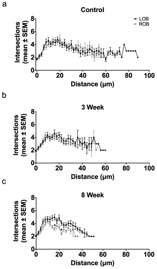
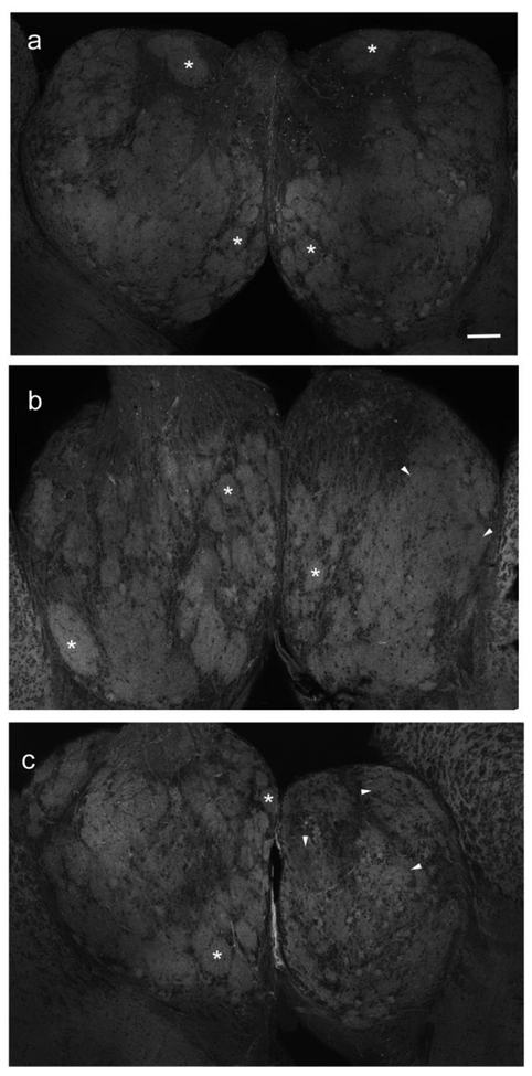
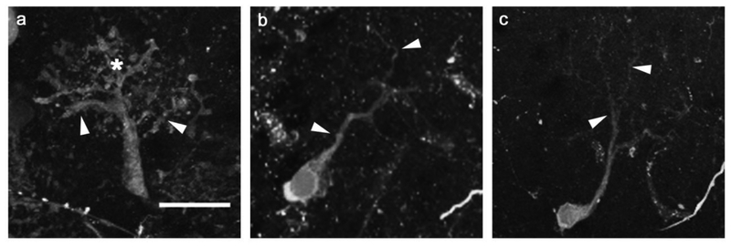
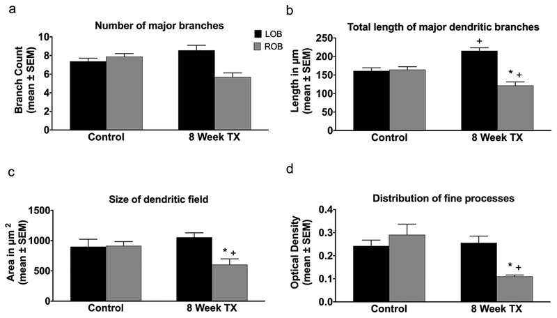
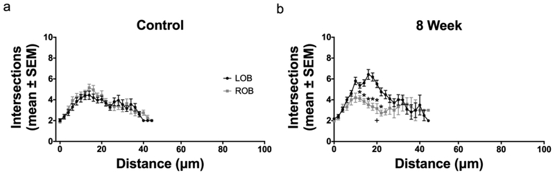
The removal of afferent input to the olfactory bulb by both cautery and chemical olfactory organ ablation in adult zebrafish results in a significant decrease in volume of the ipsilateral olfactory bulb. To examine the effects of deafferentation at a cellular level, primary output neurons of the olfactory bulb, the mitral cells, were investigated using retrograde tract tracing with fluorescent dextran using ex vivo brain cultures. Morphological characteristics including the number of major dendritic branches, total length of dendritic branches, area of the dendritic arbor, overall dendritic complexity, and optical density of the arbor were used to determine the effects of deafferentation on mitral cell dendrites. Following 8 weeks of permanent deafferentation there were significant reductions in the total length of dendritic branches, the area of the dendritic arbor, and the density of fine processes in the dendritic tuft. With 8 weeks of chronic, partial deafferentation there were significant reductions in all parameters examined, including a modified Sholl analysis that showed significant decreases in overall dendritic complexity. These results show the plasticity of mitral cell dendritic structures in the adult brain and provide information about the response of these output neurons following the loss of sensory input in this key model system.

Keywords: Dextran; Modified Sholl analysis; Output neurons; Retrograde labeling; Teleosts.


Conflict of interest statement

Conflict of Interest

The authors declare that they have no conflict of interest.

Figures

Fig. 1
Fig. 1
Parameters used to quantify the effects of deafferentation on digital reconstructions of mitral cell dendritic arbors were (a) number of major branches (gray); (b) total length of the major dendritic branches, calculated by adding the lengths of the traced branches (gray); (c) estimate of the relative size of the dendritic arbor, by connecting the distal tips (gray) of the traced branches within the dendritic arbor; and (d) distribution of fine processes of the dendritic arbor (gray), calculated from the optical density of dextran labeling of the dendritic field projection images A modified Sholl analysis was also used. (e) In a traditional Sholl analysis, concentric rings with a set distance between them are centered over the soma and the overall complexity of a dendritic arbor is evaluated. (f) A modified Sholl analysis was developed in which concentric rings with a set distance between them are placed at the base of the dendritic arbor. Using this modified Sholl analysis, mitral cells with dendritic tufts located at various distances from the soma can be directly compared
Fig. 2
Fig. 2
Dendritic morphologies of permanently deafferented mitral cells were compared with internal control cells. At the 3-week survival time, internal control cells (a) retained control morphology, with numerous major branches off the primary dendrite terminating in a dense tuft of fine processes. Scale bar = 20μm for all. Three-week deafferented cells (b) showed no obvious variation from control cells in overall gross structure of the dendritic arbor, although fine processes appeared to be less dense. At the 8-week time point, internal control cells (c) remained control-like in structure, while deafferented cells (d) exhibited reduced dendritic arbor morphology, with diminished major branches and an absence of the dense population of fine processes typically seen in control morphologies. arrowheads = major dendritic branches, * = fine processes
Fig. 3
Fig. 3
Quantitative analysis of the effects of permanent deafferentation on mitral cell dendritic arbors. (a) There were no significant changes in the number of major dendritic branches of mitral cells following 3 and 8 weeks of cautery deafferentation compared to both internal control cells and day 0 unlesioned control fish. (b) While there were no significant changed in the length of dendritic branches 3 weeks post-deafferentation, at 8 weeks post-deafferentation there was a significant reduction in the length of dendritic branches in the deafferented bulb compared to both internal control cells and day 0 unlesioned control cells. (c) At 3 weeks post-deafferentation there was no significant effect on the size of the dendritic field, but with 8 weeks of deafferentation there was a significant decrease in the size of the dendritic field compared to both internal control arbors and day 0 unlesioned control animals. (d) There were no significant changes in the distribution of fine processes with 3 weeks of deafferentation, but with 8 weeks of deafferentation there was a significant decrease in the distribution of fine processes within the dendritic arbor compared to both internal control dendritic arbors and day 0 unlesioned control arbors. * = p<0.05 compared to internal control cells; + = p<0.05 compared to day 0 unlesioned control cells; LOB = left (internal control) olfactory bulb; ROB = right (deafferented) olfactory bulb
Fig. 4
Fig. 4
Modified Sholl analysis of dendritic arbor complexity following permanent deafferentation. (a) In unlesioned control fish there was no difference in dendritic complexity between right (ROB) and left (LOB) olfactory bulbs. (b) Following 3 weeks of deafferentation there was no significant decrease in mitral cells dendritic complexity (ROB) compared to the internal control side (LOB). (c) At 8 weeks post-deafferentation, there were significant differences (*) in the number of intersections at multiple distances from the base of the arbor in deafferented cells (ROB) compared to internal control cells (LOB), and there was a trend towards a significant decrease in overall dendritic complexity (p = 0.0832). * = p<0.05 differences at specific distance from the arbor base compared to internal control
Fig. 5
Fig. 5
Effects of permanent deafferentation on the distribution of actin filaments in olfactory bulb glomeruli. (a) Unlesioned control fish had distinct glomeruli (*) throughout both right and left olfactory bulbs. Scale bar = 100μm. (b) With 3 weeks of deafferentation, some glomeruli in the deafferented bulb (star) remained intact (*) but others appeared to lose their structural features (arrowheads). The internal control bulb continued to show typical glomerular structures (*). (c) At 8 weeks post-deafferentation, the internal control bulb had typical glomeruli (*), while the deafferented bulb (star) had lost distinct glomeruli (arrowheads)
Fig. 6
Fig. 6
The effects of chronic, partial deafferentation on mitral cell dendritic morphology were examined after 8 weeks of repeated detergent infusion. (a) Mitral cells in the internal control bulb had typical dendrite morphology, including numerous major branches (arrowheads) and a dense tuft of fine processes (*). Scale bar = 20μm for all. (b, c) With eight weeks of chronic detergent infusion, deafferented cells were greatly affected, with less elaborate overall dendritic arbor structure, reduced definition of major branches (arrowheads), and minimal fine processes
Fig. 7
Fig. 7
Quantification of the effects of chronic, partial deafferentation on mitral cell dendritic structure. (a) At 8 weeks post-deafferentation there was a significant reduction in the number of major dendritic branches in deafferented mitral cells compared to internal control cells and day 0 unlesioned control animals. (b) The length of dendritic branches of 8-week deafferented cells was significantly reduced compared to internal control and day 0 unlesioned control cells. Additionally, there was a significant increase in the length of dendritic branches of internal control cells compared to day 0 unlesioned control cells. (c) Following 8 weeks of deafferentation, there was a significant decrease in the average size of dendritic arbors of deafferented mitral cells compared to internal control cells and day 0 unlesioned control cells. (d) There was a significant reduction in the distribution of fine processes within the dendritic arbor at 8 weeks post-deafferentation compared to internal control dendritic arbors and day 0 unlesioned control arbors. * = p<0.05 compared to internal control cells; + = p<0.05 compared to day 0 unlesioned control cells. LOB = left (internal control) olfactory bulb; ROB = right (deafferented) olfactory bulb
Fig. 8
Fig. 8
A modified Sholl analysis was used to determine the effects of chronic, partial deafferentation on overall dendritic arbor complexity. (a) In day 0 unlesioned control fish there were no significant differences in the dendritic complexity between right (ROB) and left (LOB) olfactory bulbs. (b) Following 8 weeks of chronic, partial deafferentation there were significant differences in the number of intersections at multiple distances from the base of the dendritic arbor (*), and there was a significant decrease in overall dendritic complexity (+), compared to internal control cells

References

    1. Baier H, Korsching S. 1994. Olfactory glomeruli in the zebrafish form an invariant pattern and are identifiable across animals. J Neurosci. 14: 219–230 - PMC - PubMed
    1. Baker H, Kawano T, Albert V, Joh TH, Reis DJ, Margolis FL. 1984. Olfactory bulb dopamine neurons survive deafferentation-induced loss of tyrosine hydroxylase. Neurosci. 11: 605–615 - PubMed
    1. Baker H, Morel K, Stone DM, Maruniak JA. 1993. Adult naris closure profoundly reduces tyrosine hydroxylase expression in mouse olfactory bulb. Brain Res. 614: 109–116 - PubMed
    1. Bloomfield SA, Hitchcock PF. 1991. Dendritic arbors of large-field ganglion cells show scaled growth during expansion of the goldfish retina: a study of morphometric and electronic properties. J Neurosci. 11: 910–917 - PMC - PubMed
    1. Braubach OR, Fine A, Croll RP. 2012. Distribution and functional organization of glomeruli in the olfactory bulbs of zebrafish (Danio rerio). J Comp Neuro. 520:2317–2339 - PubMed

Publication types

LinkOut - more resources