Review
. 2019 Feb 1;40(1):236-267.
doi: 10.1210/er.2018-00066.

Insulin-like Growth Factor-I Receptor and Thyroid-Associated Ophthalmopathy

Affiliations
Review

Insulin-like Growth Factor-I Receptor and Thyroid-Associated Ophthalmopathy

Terry J Smith et al. Endocr Rev. .

Abstract

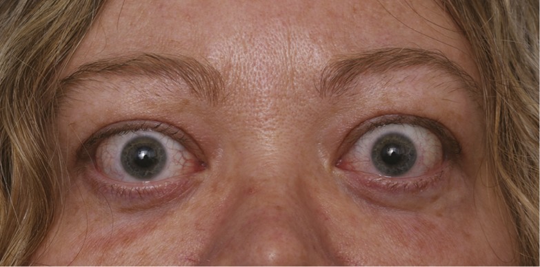
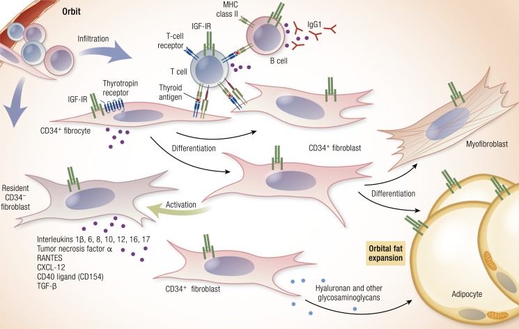
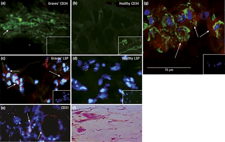
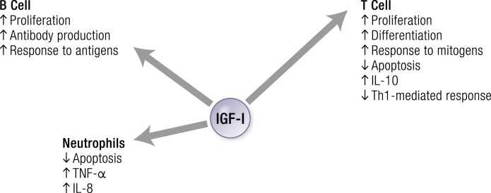
Thyroid-associated ophthalmopathy (TAO) is a complex disease process presumed to emerge from autoimmunity occurring in the thyroid gland, most frequently in Graves disease (GD). It is disfiguring and potentially blinding, culminating in orbital tissue remodeling and disruption of function of structures adjacent to the eye. There are currently no medical therapies proven capable of altering the clinical outcome of TAO in randomized, placebo-controlled multicenter trials. The orbital fibroblast represents the central target for immune reactivity. Recent identification of fibroblasts that putatively originate in the bone marrow as monocyte progenitors provides a plausible explanation for why antigens, the expressions of which were once considered restricted to the thyroid, are detected in the TAO orbit. These cells, known as fibrocytes, express relatively high levels of functional TSH receptor (TSHR) through which they can be activated by TSH and the GD-specific pathogenic antibodies that underpin thyroid overactivity. Fibrocytes also express insulin-like growth factor I receptor (IGF-IR) with which TSHR forms a physical and functional signaling complex. Notably, inhibition of IGF-IR activity results in the attenuation of signaling initiated at either receptor. Some studies suggest that IGF-IR-activating antibodies are generated in GD, whereas others refute this concept. These observations served as the rationale for implementing a recently completed therapeutic trial of teprotumumab, a monoclonal inhibitory antibody targeting IGF-IR in TAO. Results of that trial in active, moderate to severe disease revealed dramatic and rapid reductions in disease activity and severity. The targeting of IGF-IR with specific biologic agents may represent a paradigm shift in the therapy of TAO.


Figures

Figure 1.
Figure 1.
Image of patient with TAO. [© 2019 Illustration Presentation ENDOCRINE SOCIETY].
Figure 2.
Figure 2.
Theoretical schematic of the pathogenesis of TAO. Infiltrating CD34+ fibrocytes emanating from the monocyte lineage in bone marrow enter from the circulation and set the stage for disease. They express and present autoantigens reminiscent of those found in the thyroid gland. Among these are low levels of TSHR, thyroglobulin, and other thyroid antigens. Fibrocytes present antigens to antigen-specific T cells, which in turn, endorse the production of IgG1 by B cells. These fibrocytes differentiate into CD34+ fibroblasts that can differentiate further into myofibroblasts and adipocytes, depending on the signals they receive. CD34+ fibroblasts encounter residential CD34 fibroblasts. When activated, these cells generate many proinflammatory and anti-inflammatory factors, including cytokines. Among these are IL-1β, -6, -8, -10, -12, and -16; TNF-α; the chemokine known as “regulated on activation, normal T expressed and secreted” (or RANTES), CXCL12, and CD40 ligand (CD40L; CD154). These cytokines can act locally on virtually all of the cellular inhabitants of the TAO orbit. Virtually all of the infiltrating and residential cell types display IGF-IR. Thus, therapies based on the inhibition of IGF-IR might target any and all of these cells. Cytokine-activated fibroblasts synthesize hyaluronan (HA) and other glycosaminoglycans, which expand orbital tissue volume and can lead to development of proptosis. In TAO, orbital fat can expand as a likely consequence of adipogenesis. [Reproduced with permission from Smith TJ, Hegedus L. Graves’ disease. N Engl J Med. 2016;375(16):1552–1565. Copyright Massachusetts Medical Society; © 2019 Illustration Presentation ENDOCRINE SOCIETY].
Figure 3.
Figure 3.
CD34+ fibrocytes infiltrate the orbit in TAO. OFs derived from patients with TAO comprise discrete subsets of cells on the basis of CD34 display. CD34+lymphocyte-specific protein (LSP) 1+TSHR+ fibrocytes are present in TAO orbital tissue but not in healthy tissues. (a) CD34 expression (arrows) in TAO-derived tissue (inset, negative control). (b) Undetectable CD34 in healthy tissue (inset, positive control). (c) LSP-1 expression in TAO tissue [red; arrows (inset, negative control)]. (d) Undetectable LSP-1 in healthy tissue (inset, negative control). (e) CD31 expression in TAO tissue is limited to vascular endothelium (red; arrows). (f) Hematoxylin and eosin-stained, consecutive thin sections of the same orbital tissue. (g) Fibrocytes present in TAO orbital tissue coexpress CD34 and TSHR. Arrows denote fibrocytes. [Reproduced with permission from Douglas RS, Afifiyan NF, Hwang CJ, Chong K, Haider U, Richards P, Gianoukakis AG, Smith TJ. Increased generation of fibrocytes in thyroid-associated ophthalmopathy. J Clin Endocrinol Metab. 2010;95(1):430–438; © 2019 Illustration Presentation ENDOCRINE SOCIETY].
Figure 4.
Figure 4.
Fibrocytes express several proteins thought to be restricted to the thyroid. Expression of the proteins appears to depend on autoimmune regulator protein (AIRE). Fibrocytes collected from an individual with autoimmune polyendocrine syndrome type 1 (APS-1) express lower levels of AIRE, TSHR, thyroglobulin (Tg), sodium iodide symporter (NIS), thyroid peroxidase (TPO), pair-boxed 8 (PAX8), and thyroid transcription factor-1 (TTF-1) than do those from an unaffected, first-degree relative. Results from real-time PCR of the targets are indicated. [Reproduced with permission from Fernando R, Lu Y, Atkins SJ, Mester T, Branham K, Smith TJ. Expression of TSHR, thyroglobulin, sodium iodide symporter, and thyroteroxidase by fibrocytes depends on AIRE. J Clin Endocrinol Metab. 2014;99(7):E1236–E1244; © 2019 Illustration Presentation ENDOCRINE SOCIETY].
Figure 5.
Figure 5.
Overview of the IGF-I pathway. IGF-I, IGF-II, insulin, IGF-binding proteins (IGFBPs), IGF-IR, IGF-IIR, and insulin receptor (IR) form a complex pathway. IGF-IR predominantly regulates growth, whereas the IR dominates regulation of metabolism. IGF-IIR (mannose-6-phosphate receptor) regulates IGF-II degradation. IGF-I and IGF-II action is modulated by six IGFBPs. ALS, acid-labile subunit. [Adapted with permission from Lowe WL Jr. Insulin-like growth factors. Sci Med. 1996;3(2):62–71; © 2019 Illustration Presentation ENDOCRINE SOCIETY].
Figure 6.
Figure 6.
IGF-IR, IGF-IIR, and IR are displayed on the cell surface. IGF-IR and IGF-IR/IR hybrid proteins are preferentially activated by IGF-I rather than insulin. [Adapted with permission from Ryan PD, Goss PE. The emerging role of the insulin-like growth factor pathway as a therapeutic target in cancer. Oncologist. 2008;13(1):16–24; © 2019 Illustration Presentation ENDOCRINE SOCIETY].
Figure 7.
Figure 7.
Classical model of signaling through the IGF-IR. This results in downstream canonical (MAPK)/Ras-Raf-Erk and phosphatidylinositol 3-kinase (PI3K)/AKT/mammalian target of rapamycin (mTOR) phosphorylation. IRS, IR substrate; MEK, MAPK kinase; P, phosphorylation. [Adapted from Worrall C, Nedelcu D, Serly J, Suleymanova N, Oprea I, Girnita A, Girnita L. Novel mechanisms of regulation of IGF-1R action: functional and therapeutic implications. Pediatr Endocrinol Rev. 2013;10(4):473–484; © 2019 Illustration Presentation ENDOCRINE SOCIETY].
Figure 8.
Figure 8.
The IGF-IR is now considered a functional RTK/GPCR hybrid, integrating canonical tyrosine kinase and GPCR signaling. Ligand binding activates classical kinase-dependent signaling pathways, as well as β-arrestin recruitment to GPCR kinase-dependent, phosphorylated serine residues within the C-tail of IGF-IR. β-Arrestin (β-ARR1) induces kinase desensitization and receptor ubiquitination while provoking kinase-independent signaling through the MAPK pathway. [Adapted from Worrall C, Nedelcu D, Serly J, Suleymanova N, Oprea I, Girnita A, Girnita L. Novel mechanisms of regulation of IGF-1R action: functional and therapeutic implications. Pediatr Endocrinol Rev. 2013;10(4):473–484; © 2019 Illustration Presentation ENDOCRINE SOCIETY].
Figure 9.
Figure 9.
IGF-I exerts regulatory actions on immune function by stimulating B cells, T cells, and neutrophils. [© 2019 Illustration Presentation ENDOCRINE SOCIETY].
Figure 10.
Figure 10.
Evidence for the physical association of IGF-IR and TSHR in orbital tissues in situ, cultured OFs, and thyroid epithelial cells in vitro. (a–f) Immunofluorescence staining using an anti–IGF-IRβ antibody appearing as red and anti-TSHR antibody appearing as green by confocal microscopy. (a–c) OFs from a patient with TAO (GD-OFs). (d–f) Primary human thyrocytes. (g–i) Another set of antibodies is used where IGF-IRβ appears as green and TSHR as red. (j–l) TSHR is green, and IGF-IRα is red in GD-OFs. (m–o) TAO orbital tissue where TSHR is green, and IGF-IR is red. (c, f, i, l, and o) Merged images are captured. [Reproduced with permission from Tsui S, Naik V, Hoa N, Hwang CJ, Afifiyan NF, Sinha Hikim A, Gianoukakis AG, Douglas RS, Smith TJ. Evidence for an association between thyroid-stimulating hormone and insulin-like growth factor 1 receptors: a tale of two antigens implicated in Graves' disease. J Immunol. 2008;181(6):4397–4405. Copyright 2008 The American Association of Immunologists, Inc; © 2019 Illustration Presentation ENDOCRINE SOCIETY].
Figure 11.
Figure 11.
Evidence for specific activating antibodies recognizing IGF-IR in patients with GD. Expression of a dominant-negative (DN) mutant IGF-IR transfected into GD-OF blocks the induction by GD-IgG of (a) IL-16- and RANTES-dependent T cell chemoattraction and (b) protein expression. [Reproduced with permission from Pritchard J, Han R, Horst N, Cruikshank WW, Smith TJ. Immunoglobulin activation of T cell chemoattractant expression in fibroblasts from patients with Graves’ disease is mediated through the insulin-like growth factor I receptor pathway. J Immunol. 2003;170(12):6348–6354. Copyright 2003 The American Association of Immunologists, Inc.; © 2019 Illustration Presentation ENDOCRINE SOCIETY].
Figure 12.
Figure 12.
Screening, randomization, response, and follow-up of patients’ participation in clinical trial RV001. (a) Patients meeting inclusion criteria entered the trial-screening process. At baseline, patients were randomized to receive active drug or placebo for the 24-week intervention phase. This was followed by a 1-year observation. (b) An analysis to first response. (c) The time course of patients meeting response criteria. (d) Responses are graded at week 24, where a high response indicates ≥3 mm proptosis and clinical activity score (CAS) reduced greater than or equal to three points on a seven-point scale. [Reproduced with permission from Smith TJ, Kahaly GJ, Ezra DG, Fleming JC, Dailey RA, Tang RA, Harris GJ, Antonelli A, Salvi M, Goldberg RA, Gigantelli JW, Couch SM, Shriver EM, Hayek BR, Hink EM, Woodward RM, Gabriel K, Magni G, Douglas RS. Teprotumumab for thyroid-associated ophthalmopathy. N Engl J Med. 2017;376(18):1748–1761. Copyright Massachusetts Medical Society; © 2019 Illustration Presentation ENDOCRINE SOCIETY].
Figure 13.
Figure 13.
Secondary efficacy end points in clinical trial RV001. (a) The time course of changes in proptosis from baseline. (b) The time course of change from baseline in CAS. (c) Post hoc analysis of the fraction of patients with CAS of zero or one at the time point indicated along the abscissa. (c) Change in visual functioning subscale of the QOL scale (GO-QOL). (d) GO-QOL visual-functioning subscale. (e) Change in appearance subscale of GO-QOL. (f) Responses in terms of diplopia. [Reproduced with permission from Smith TJ, Kahaly GJ, Ezra DG, Fleming JC, Dailey RA, Tang RA, Harris GJ, Antonelli A, Salvi M, Goldberg RA, Gigantelli JW, Couch SM, Shriver EM, Hayek BR, Hink EM, Woodward RM, Gabriel K, Magni G, Douglas RS. Teprotumumab for thyroid-associated ophthalmopathy. N Engl J Med. 2017;376(18):1748–1761. Copyright Massachusetts Medical Society; © 2019 Illustration Presentation ENDOCRINE SOCIETY].

Similar articles

Cited by

References

    1. Smith TJ, Hegedüs L. Graves’ disease. N Engl J Med. 2016;375(16):1552–1565. - PubMed
    1. Nordyke RA, Gilbert FI Jr, Harada AS. Graves’ disease. Influence of age on clinical findings. Arch Intern Med. 1988;148(3):626–631. - PubMed
    1. Bartalena L, Baldeschi L, Boboridis K, Eckstein A, Kahaly GJ, Marcocci C, Perros P, Salvi M, Wiersinga WM; European Group on Graves’ Orbitopathy (EUGOGO) . The 2016 European Thyroid Association/European Group on Graves’ Orbitopathy Guidelines for the Management of Graves’ Orbitopathy. Eur Thyroid J. 2016;5(1):9–26. - PMC - PubMed
    1. Chan W, Wong GW, Fan DS, Cheng AC, Lam DS, Ng JS. Ophthalmopathy in childhood Graves’ disease. Br J Ophthalmol. 2002;86(7):740–742. - PMC - PubMed
    1. Putta-Manohar S, Perros P. Epidemiology of Graves’ orbitopathy. Pediatr Endocrinol Rev. 2010;7(Suppl 2):182–185. - PubMed

Publication types

MeSH terms