. 2018 Nov 1;28(11):4105-4119.
doi: 10.1093/cercor/bhy225.

Inhibitory Neuron Activity Contributions to Hemodynamic Responses and Metabolic Load Examined Using an Inhibitory Optogenetic Mouse Model

Affiliations

Inhibitory Neuron Activity Contributions to Hemodynamic Responses and Metabolic Load Examined Using an Inhibitory Optogenetic Mouse Model

Alberto L Vazquez et al. Cereb Cortex. .

Abstract

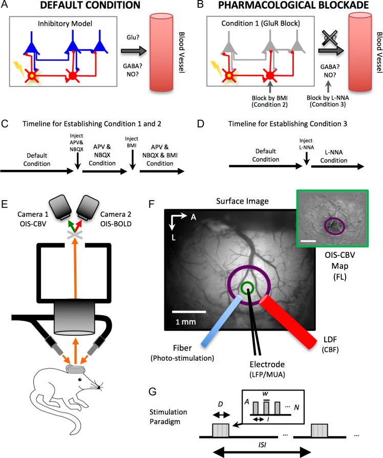
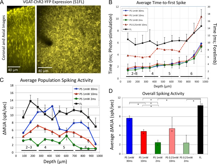
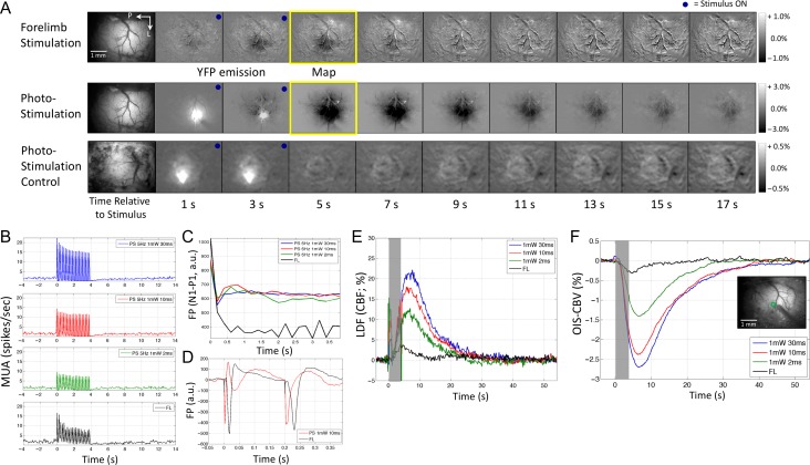
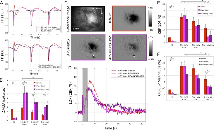
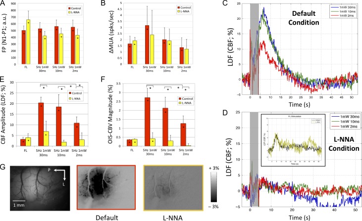
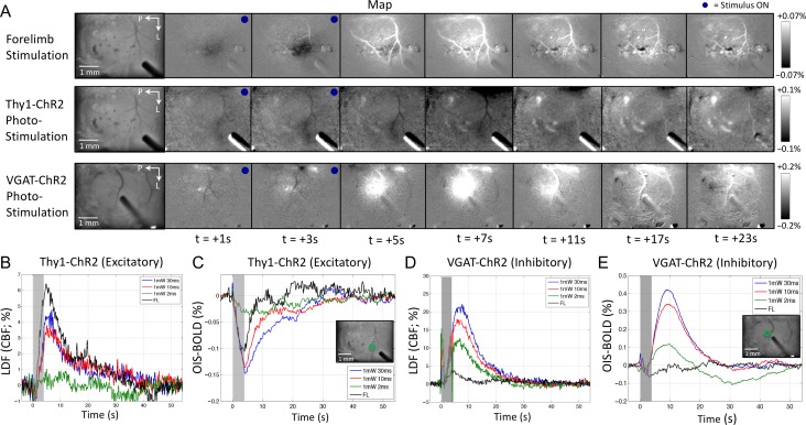
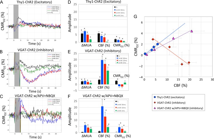
Hemodynamic signals are routinely used to noninvasively assess brain function in humans and animals. This work examined the contribution of inhibitory neuron activity on hemodynamic responses captured by changes in blood flow, volume and oxygenation in the cortex of lightly anesthetized mice. Because cortical activity is not commonly initiated by inhibitory neurons, experiments were conducted to examine the neuronal activity properties elicited by photo-stimulation. We observed comparable increases in neuronal activity evoked by forelimb and photo-stimulation; however, significantly larger increases in blood flow and volume were produced by photo-stimulation of inhibitory neurons compared with forelimb stimulation. Following blockade of glutamate and GABA-A receptors to reduce postsynaptic activity contributions, neuronal activity was reliably modulated and hemodynamic changes persisted, though slightly reduced. More importantly, photo-stimulation-evoked changes in blood flow and volume were suppressed by 75-80% with the administration of a nitric oxide synthase inhibitor, suggesting that inhibitory neurons regulate blood flow mostly via nitric oxide. Lastly, forelimb and photo-stimulation of excitatory neurons produced local decreases in blood oxygenation, while large increases were generated by photo-stimulation of inhibitory neurons. Estimates of oxygen metabolism suggest that inhibitory neuron activity has a small impact on tissue metabolic load, indicating a mismatch between the metabolic demand and blood flow regulation properties of inhibitory and excitatory neurons.


Figures

Figure 1.
Figure 1.
Simplified schematic of the experimental conditions implemented in this work and experimental setup. (A) Under default conditions (light ketamine anesthesia), photo-stimulation activates inhibitory neurons (yellow soma) which will likely inhibit postsynaptic excitatory (glutamatergic in blue) and inhibitory (gabaergic in red) neurons. Some inhibitory neurons are known to release nitric oxide (NO), a potent vasodilator. (B) Experiments were also performed under pharmacological blockade of glutamate receptor (GluR) activity to prevent the modulation of excitatory activity (Condition 1). Pharmacological blockade of GABAA receptors was also used to prevent fast inhibitory action on inhibitory (and excitatory) neurons (Condition 2). Lastly, experiments were performed under pharmacological blockade of nitric oxide synthase (NOS) to determine the role nitric oxide on blood flow regulation by inhibitory neuron activity (Condition 3). (C) Timeline for establishing Conditions 1 and 2. These conditions were established in sequence. Experiments under a combination of APV, NBQX, and BMI were conducted while monitoring background ongoing activity to ensure this condition is maintained during data collection. (D) Timeline for establishing Condition 3. Experimental setup. (E) Optical images of intrinsic signal (OIS) were acquired simultaneously from the somatosensory cortex of lightly anesthetized mice using a dual camera system. One camera acquired images sensitive to changes in cerebral blood volume (OIS–CBV) and the other camera acquired images sensitive to changes in blood oxygenation (OIS–BOLD). The brain was illuminated with orange light and appropriately filtered prior to each camera. (F) Probe placement and ROI. An initial mapping experiment was used to map the location of the forelimb somatosensory cortex for further experimentation (top-right inset; scale bar = 1 mm). The optic fiber, electrode, and laser Doppler flowmetry (LDF) probe were placed in the center of the activated region (purple circle) while avoiding large surface vasculature. The ROI used to obtain OIS time series (OIS–CBV and OIS–BOLD) is shown in green, it excluded surface vasculature. (G) Stimulation paradigm. Forelimb or photo-stimuli were delivered with amplitude A, pulse duration w, at a frequency or interval I, over an overall stimulus duration D. These stimulation trials were repeated after time period ISI.
Figure 2.
Figure 2.
Characterization of the optogenetic inhibitory mouse model. (A) Two-photon microscopy images of ChR2-YFP expression in the somatosensory cortex. Images of an orthogonal section along depth (coronal section on the left) and axial section 200 μm under the cortical surface show the numerous cells expressing ChR2 in the somatosensory cortex. (B) Average time-to-first spike after evoked photo-stimulation (left y-axis) for different stimulation parameters and after forelimb stimulation (black line, right y-axis) as a function of depth. The approximate locations of the cortical layers are shown along the bottom of the horizontal axis (Altamura et al. 2007). (C) Average change in multiunit activity (ΔMUA) as a function of depth for different photo-stimulation parameters as well as forelimb stimulation (black line). (D) Average overall spiking activity for all photo-stimulation parameters tested. On average, forelimb activity was largest though not significantly larger than that evoked by 1 mW 30 ms photo-stimulation. Error bars denote the standard error and statistically significant differences (P < 0.05) are denoted by the asterisk in the bar (significantly greater than 0) or the lines above the bar graph for population comparisons.
Figure 3.
Figure 3.
Neural and hemodynamic responses obtained under default conditions for Experiment 2. (A) Maps of the spatiotemporal evolution of hemodynamic responses (OIS–CBV) evoked by forelimb stimulation (top row) and photo-stimulation (10 ms pulse duration; middle row) in this mouse model (VGAT-ChR2). Maps from photo-stimulation of a control model (expressing GFP but not ChR2) are also shown (bottom row; 1 mW 30 ms pulses). The stimulation period lasted 4 s and is denoted by the blue dot on the top-right corner of each frame. Increases in blood volume absorb more light and are therefore manifested by darkening of the image. Photo-stimulation also excites YFP coexpressed with ChR2 in this mouse model (or GFP in the control model), hence, the brightened area showing the photo-stimulated location. (B) Average changes in multiunit activity (MUA) in response to photo-stimulation and forelimb stimulation (black line, bottom-most panel). (C) Average field potential (FP) response envelope during the stimulation period for both modes of stimulation. (D) Average FP trace showing the average changes in field potential of photo-stimulation (10 ms pulses, red line, top panel) and forelimb stimulation (black line, bottom panel). Small electrical stimulation artifacts are present for each delivered stimulus (5 Hz frequency or every 200 ms; first 2 pulses shown). (E) Average changes in cerebral blood flow (CBF) evoked by photo-stimulation or forelimb stimulation measured by laser Doppler flowmetry (LDF). The stimulation period is denoted by the gray bar. The rapid LDF increase with photo-stimulation onset is artefactual and due to interference between the LDF flowmeter and the photo-stimulation laser. (F) Average changes in OIS–CBV signal. Decreases in OIS–CBV correspond to increases in CBV. Inset shows a sample OIS–CBV image with the ROI location (green circle) for time series extraction.
Figure 4.
Figure 4.
Neural and hemodynamic responses obtained under default (red), APV+NBQX (pink), and APV+NBQX+BMI (purple) conditions for Experiment 3. (A) Average field potential (FP) waveform for the first 3 forelimb (FL) and photo-stimulation pulses (10 ms pulse duration shown). (B) Average change in multiunit activity (∆MUA) evoked by forelimb and photo-stimulation. (C) OIS–CBV maps obtained from a sample subject immediately after photo-stimulation under default (red border), APV + NBQX (pink border) and APV+NBQX+BMI conditions (purple border). Map scale bar is shown to the left of each row. (D) Average blood flow changes evoked by photo-stimulation (10 ms pulse duration shown) under the 3 conditions tested in Experiments 2 and 3. The gray bar denotes the photo-stimulation period. The initial rapid change with photo-stimulation onset and offset are artefactual and due to interference between the LDF flowmeter and photo-stimulus. Average change in CBF amplitude (E) and OIS–CBV magnitude (F) evoked by forelimb and photo-stimulation. Error bars denote standard error. Statistically significant differences (P < 0.05) are denoted by the asterisk in the bar (significantly greater than 0) or the lines above the bar graph for population comparisons. Significant comparisons between forelimb and photo-stimulation are not included in panels B, E, or F for visibility, most are significantly different, especially those from Experiment 2 and 3.
Figure 5.
Figure 5.
Neural and hemodynamic responses obtained under default and L-NNA conditions (Experiment 4). (A) Average field potential (FP) waveform evoked by photo-stimulation (10 ms pulse duration shown; top panel) and forelimb stimulation (bottom panel). (B) Average change in multiunit activity (∆MUA), (E) CBF amplitude and (F) OIS–CBV magnitude for the different stimulation parameters. (C) Average changes in blood flow to the different photo-stimuli tested under default conditions and (D) after the intracortical administration of the NOS blocker L-NNA (0.5 mM). In general, small changes in blood flow were observed. The inset in D shows the changes in blood flow evoked by forelimb stimulation before and after L-NNA administration. (G) OIS–CBV maps produced by photo-stimulation (10 ms shown) in a sample subject. Error bars denote standard error. Statistically significant differences (P < 0.05) are denoted by the asterisk in the bar (significantly greater than 0) or the lines above the bar graph for population comparisons. Significant comparisons between forelimb and photo-stimulation are not included in panels A, B, E, or F to reduce clutter.
Figure 6.
Figure 6.
Changes in OIS–BOLD signal evoked by forelimb and photo-stimulation in excitatory and inhibitory optogenetic mouse models. (A) Example of spatiotemporal changes in OIS–BOLD signal in response to forelimb (FL, 5 Hz, top row panel) and photo-stimulation (PS) from one Thy1-ChR2 mouse (excitatory optogenetic model; middle row panel; 1 mW, 30 ms pulses at 5 Hz shown). Example of the spatiotemporal changes in OIS–BOLD signal evoked by photo-stimulation from one VGAT-ChR2 mouse (inhibitory optogenetic model; 1 mW, 10 ms pulses delivered at 5 Hz; bottom row panel). Each image sequence shows the baseline image on the left, followed by images of the changes in signal relative to baseline. Each frame shows the average change over a 1-s time span. The temporal sequence relative to stimulation onset is indicated below the bottom-most panel sequence in A. Evident decreases in OIS–BOLD signal are observed in parenchymal regions followed by increases especially in the venous vasculature and sinus to forelimb stimulation in Thy1-ChR2 mice. Mostly decreases in OIS–BOLD signal are observed in response to photo-stimulation of Thy1-ChR2 mice. On the other hand, large increases in OIS–BOLD signal are observed in parenchymal regions as well as venous vasculature as a result of photo-stimulation in VGAT-ChR2 mice. Decreasing the color scale extent did not reveal parenchymal decreases in OIS–BOLD signal in these mice. Average changes in blood flow and OIS–BOLD over the selected ROI (circular region centered on the insertion location of the electrode excluding surface vasculature) produced by forelimb and photo-stimulation from all Thy1-ChR2 mice (panels B and C, respectively) and all VGAT-ChR2 mice (panels D and E, respectively). Inset images in panels C and E show sample locations of the ROI (inner region inside the green circle). A sample ROI location is also presented in Figure 1F (green circle). The changes in blood flow and blood oxygenation evoked by forelimb stimulation were similar in both mouse models though slightly smaller in amplitude for the inhibitory mouse model. In the excitatory model, all changes in blood oxygenation were negative indicative of stimulation-evoked increases in tissue oxygen metabolism. However, the changes in blood oxygenation observed in inhibitory mice were large and positive, suggesting that the increases in blood flow were disproportionately larger to the increases in tissue oxygen metabolism.
Figure 7.
Figure 7.
Estimates of CMRO2 changes evoked by forelimb and photo-stimulation in excitatory and inhibitory optogenetic mouse models. Average LDF, OIS–CBV and OIS–BOLD data from each animal were used to calculate changes in CMRO2 using Equation 1. Average changes in oxygen metabolism (CMRO2; in % relative to baseline) due to forelimb and photo-stimulation are shown in (A) for Thy1-ChR2 mice (excitatory optogenetic mouse model) and (B) for VGAT-ChR2 mice (inhibitory optogenetic mouse model) under default conditions as well as (C) for VGAT-ChR2 mice under APV+NBQX conditions. The gray bar denotes the stimulation period; photo-stimulation introduced some artifacts into the data especially around stimulation onset and offset, hence, average time series during stimulation may not reflect actual changes (photo-stimulation data only). Summary bar graphs of the average changes in spiking activity (∆MUA, spikes/s), changes in CBF (%) and changes in CMRO2 (%) are shown in panel D, panel E, and panel F for Thy1-ChR2 mice and VGAT-ChR2 mice under default conditions as well as VGAT-ChR2 mice under APV+ NBQX conditions, respectively. The average spike rate was obtained during the stimulation period while the average change in CBF and CMRO2 was obtained as the average change between 4 and 7 s after stimulation onset for all animals. (G) Summary scatter plot of the average changes in CBF (%, x-axis) and CMRO2 (%, y-axis) from the excitatory mouse model (blue circles) and inhibitory mouse model (red diamonds for default condition responses and purple triangle for responses obtained under APV+NBQX conditions). A linear model was fit to all the data from each animal model and slopes of 0.321 (P < 0.01, R2 = 0.67), −0.358 (P > 0.15, R2 = 0.04) and 0.218 (P < 0.01, R2 = 0.53) were calculated for the excitatory model and inhibitory model under default and APV+NBQX conditions, respectively. Error bars denote standard error. All 3 linear models were significantly different from one another (P > 0.10). Statistically significant differences are denoted by the asterisk in the bar (significantly different from 0, P < 0.05).

References

    1. Adachi K, Takahashi S, Melzer P, Campos KL, Nelson T, Kennedy C, Sokoloff L. 1994. Increases in local cerebral blood flow associated with somatosensory activation are not mediated by NO. Am J Physiol. 267:H2155–H2162. - PubMed
    1. Altamura C, Dell’Acqua ML, Moessner R, Murphy DL, Lesch KP, Persico AM. 2007. Altered neocortical cell density and layer thickness in serotonin transporter knockout mice: a quantitation study. Cereb Cortex. 17:1394–1401. - PubMed
    1. Anenberg E, Chan AW, Xie Y, LeDue JM, Murphy TH. 2015. Optogenetic stimulation of GABA neurons can decrease local neuronal activity while increasing cortical blood flow. J Cereb Blood Flow Metab. 35:1579–1586. - PMC - PubMed
    1. Armstrong-James M, Fox K, Das-Gupta A. 1992. Flow of excitation within rat barrel cortex on striking a single vibrissa. J Neurophysiol. 68:1345–1358. - PubMed
    1. Boorman L, Harris S, Bruyns-Haylett M, Kennerley A, Zheng Y, Martin C, Jones M, Redgrave P, Berwick J. 2015. Long-latency reductions in gamma power predict hemodynamic changes that underlie the negative BOLD signal. J Neurosci. 35:4641–4656. - PMC - PubMed

Publication types