Review
. 2018 Sep 14;8(4):1575-1602.
doi: 10.1002/cphy.c170033.

Neural Basis of Touch and Proprioception in Primate Cortex

Affiliations
Review

Neural Basis of Touch and Proprioception in Primate Cortex

Benoit P Delhaye et al. Compr Physiol. .

Abstract

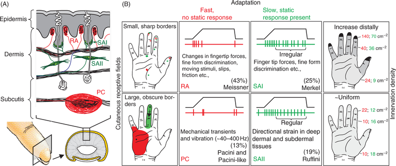
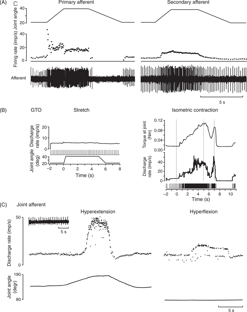
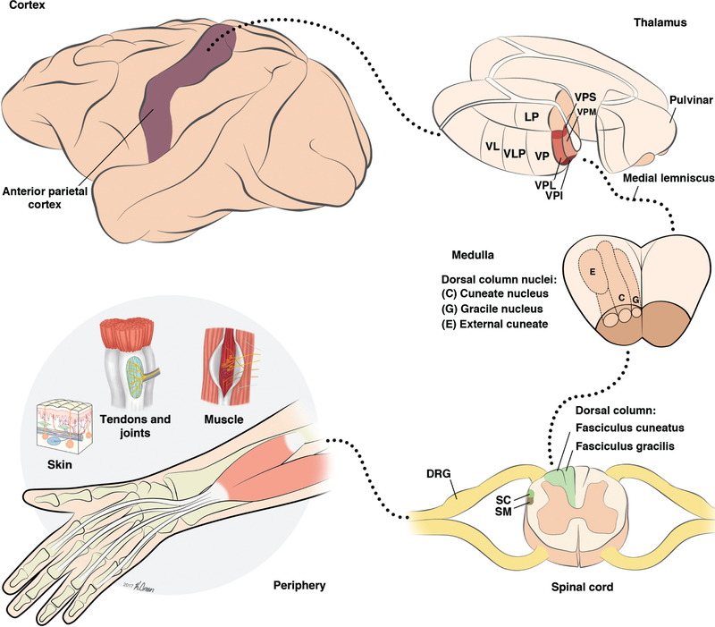
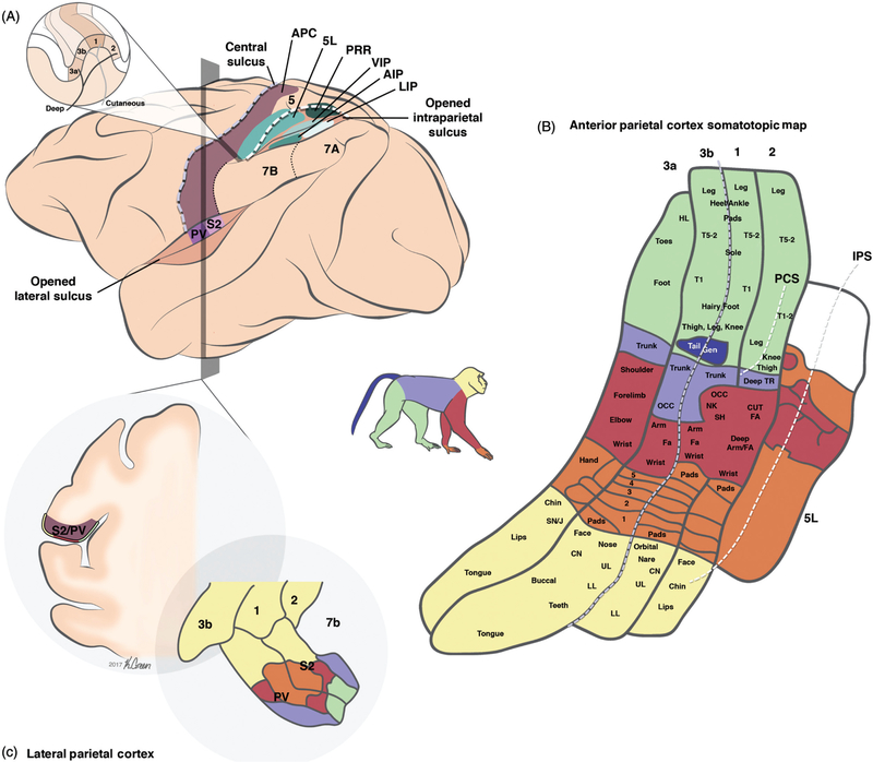
The sense of proprioception allows us to keep track of our limb posture and movements and the sense of touch provides us with information about objects with which we come into contact. In both senses, mechanoreceptors convert the deformation of tissues-skin, muscles, tendons, ligaments, or joints-into neural signals. Tactile and proprioceptive signals are then relayed by the peripheral nerves to the central nervous system, where they are processed to give rise to percepts of objects and of the state of our body. In this review, we first examine briefly the receptors that mediate touch and proprioception, their associated nerve fibers, and pathways they follow to the cerebral cortex. We then provide an overview of the different cortical areas that process tactile and proprioceptive information. Next, we discuss how various features of objects-their shape, motion, and texture, for example-are encoded in the various cortical fields, and the susceptibility of these neural codes to attention and other forms of higher-order modulation. Finally, we summarize recent efforts to restore the senses of touch and proprioception by electrically stimulating somatosensory cortex. © 2018 American Physiological Society. Compr Physiol 8:1575-1602, 2018.


Figures

Figure 1
Figure 1
The four classes of cutanueous afferents of the glabrous skin. (A) Morphology of the different mechanoreceptors and their respective locations in the skin. (B) Adaptation properties and receptive field (RF) size of the four classes of cutaneous afferents. Rapidly adapting (sometimes referred to as fast adapting, particularly for humans) versus slowly adapting refers to responses to indentations (transient vs. sustained, respectively). Type I versus type II refers to the size of the RFs, determined in part by the depth of the mechanoreceptors in the skin: Type I fibers have small RFs whereas type II fibers have large ones. The density of innervation depends on the fiber type: Type I fibers innervate the skin more densely than do type II fibers. For example, rapidly adapting afferent type II (PC) afferents show rapidly adapting responses with large RFs and relatively low innervation density (type II). Adapted, with permission, from (183).
Figure 2
Figure 2
Typical responses of proprioceptive afferents. (A) Responses of a primary (left) and secondary (right) spindle afferent from the finger extensors muscles to passive ramp and hold stretches applied to the metacarpophalangeal (MCP) joint. Primary afferents tend to be more sensitive to changes in length than secondary ones. Adapted, with permission, from (95). (B) Golgi tendon organ (GTO) do not respond to passive ramp and hold stretches (left) but respond robustly to isometric contraction (right). Adapted, with permission, from (96). (C) Responses of a joint afferent associated with the proximal interphalangeal joint of the index finger during passive manipulations. Joint receptors tend to only respond at the extrema of joint movements, perhaps to signal the threat of injury. Adapted, with permission, from (30).
Figure 3
Figure 3
Pathways from somatosensory periphery to cortex. Afferent fibers at the periphery bundle in fascicles that join to form the nerves. Afferent cell bodies are gathered in the dorsal root ganglia (DRG). When entering the spinal cord through the dorsal root, afferent axons branch, sending one projection to the dorsal horn and one projection to the dorsal column nuclei (DCN) through the dorsal column. The DCN projects contralaterally through the medial lemniscus to the ventroposterior complex of the thalamus, which in turns relays the information to cortex. Abbreviations: Dorsal root ganglion (DRG); spinomedullothalamic (SM), and spinocervicothalamic (SC) tracts. Thalamus: ventral posterior (VP), posterolateral (VPL), posteromedial (VPM), posterior inferior (VPI) and posterior superior (VPS) nuclei, posterior division (VLp) of the ventral lateral nucleus (VL), lateral posterior nucleus (LP).
Figure 4
Figure 4
Organization of somatosensory cortical areas. (A) A lateral view of the brain showing the different somatosensory areas in macaque monkey cortex. Adapted, with permission, from (198). Inset: Horizontal section of the postcentral gyrus at the level of the hand representation, showing the position of the different APC modules relative to the central and the intraparietal sulci. (B) Detailed view of the somatotopic representation of the body in the four fields of APC (areas 3a, 3b, 1, and 2) and in area 5L. Adapted, with permission, from (261, 332). (C) Coronal section showing the location of LPC in the lateral sulcus. Adapted, with permission, from (214). Abbreviations: Anterior parietal cortex (APC); second somatosensory area (S2); parietal ventral area (PV); parietal reaching region (PRR); anterior (AIP), ventral (VIP) and lateral (LIP) intraparietal areas; post central sulcus (PCS); intraparietal sulcus (IPS). Somatotopic map: Upper lip (UL); lower lip (LL); chin (CN); snout/jaw (SN/J); digits of the hand (–5); (cutaneous) forearm ((CUT) FA); occiput (OCC); trunk (TR); toes (T1–5); hindlimb (HL).
Figure 5
Figure 5
Major connections between somatosensory areas. Schematic representation of the major connections between somatosensory areas in the central nervous system, split into four major regions: the thalamus, the anterior parietal cortex (APC), the lateral parietal cortex (LPC), and the posterior parietal cortex (PPC). Abbreviations: Ventral posterior nucleus (VP), anterior pulvinar nucleus (Pla), secondary somatoensory cortex (S2), parietal ventral area (PV), parietal reach region (PRR). Area 5 also receive input from the lateral posterior nucleus in thalamus (LP, not shown in the chart).
Figure 6
Figure 6
Submodality convergence in APC. (A) Trajectory of a punctate stimulus indented 2 mm into the center of a neuron’s receptive field. (B) Typical response of a slowly adapting type 1 (SA1) and rapidly adapting (RA) afferent to 60 repeated presentations of the stimulus. (C) Response of typical neurons in area 3b. Some neurons respond throughout the stimulation interval and do not show phasic off responses, similar to SA1 fibers; others respond with phasic on-off responses, similar to RA1 fibers, but the majority respond with a mixture of sustained and phasic responses, implying input from both fiber types. Adapted, with permission, from (274).
Figure 7
Figure 7
Spatial structure of receptive fields of a neuron in area 3b. The two squares in each group display the RF estimated from the raw data (left) and the positions of the modeled Gaussian representations (right). Left: The experimental RF was obtained by continuously scanning the finger with a random pattern of raised dots, and then computing an RF map using reverse correlation (see (82)). Dark regions are excitatory, white regions are inhibitory. Right: The locations of the excitatory (solid ellipse) and fixed inhibitory components are unaffected by scanning direction and the lagged inhibitory component (dotted ellipse) trails the center by a fixed distance in each direction. Reproduced, with permission, from (81).
Figure 8
Figure 8
In contrast to their counterparts in primary somatosensory cortex, neurons in secondary somatosensory cortex exhibit task- dependent modulation in their responses to identical vibratory stimuli. (A) Spiking responses recorded from one neuron in APC and one in LPC (adapted, with permission, from (152) and from (311), respectively). Each row shows the response to a pair of stimuli: 10 or 26 Hz in the first stimulus period (f1) and 18 Hz in the second (f2). In APC, the response to f2 is independent of f1, while in LPC, the response to f2 is greater when f2 > f1. (B) LPC firing rates as a function of the frequency of vibration in the tactile stimulus. During the first stimulation period (f1), rates decrease monotonically as stimulus frequency increases. During the comparison period (f2), neurons respond preferentially when f2 > f1 (shown here) or vice versa (black points show trials on which f2 > f1; green traces denote trials on which f2 < f1).
Figure 9
Figure 9
Temporal patterning in peripheral and cortical responses to sinusoidal vibrations applied to the skin. (A) Typical response of peripheral afferents (two SAI in green, two RA in blue, and two PC in orange) to sinusoidal vibrations (amplitude = 250 microns) of different frequencies applied in the center of their RF. The responses of tactile fibers are strongly phase-locked to the stimulus and highly repeatable. Data adapted, with permission, from (252). (B) Responses of two typical APC neurons to sinusoidal vibrations. APC neurons show various degrees of phase-locking and greater trial-to-trial variability. Within this low-frequency range, the frequency of the stimulus can be extracted from both the temporal patterning of the response and the mean firing rate. Reproduced, with permission, from (152, 314).
Figure 10
Figure 10
Spatial processing in the somatosensory system. (A) Reconstructed response of tactile nerve fibers to embossed letters scanned across the skin. As in the retina, the spatial configuration of the stimulus is reflected in the spatial pattern of activation it evokes in SA1 and RA populations. Reproduced, with permission, from (282). (A) Responses of a neuron in area 3b to oriented edges indented into the skin (eight orientations, three indentation depths). This neuron is strongly tuned for edge orientation, as are neurons in primary visual cortex. Reproduced, with permission, from (11). (C) Responses of an LPC neuron to curvatures indented into the skin. This neuron prefers intermediate curvatures with the convex end pointing proximally. This type of feature selectivity is not observed in early stages of cortical processing (e.g., in area 3b). Reproduced, with permission, from (391). (D) Responses of an LPC neuron to bars indented into the skin. This neuron exhibits the similar preferred orientation over large swaths of skin (position-invariant orientation tuning). Reproduced, with permission, from (107).
Figure 11
Figure 11
Motion coding in APC. (A) Direction tuning of a neuron in area 3b to bars scanned across its receptive field. Adapted, with permission, from (276). (B) The geometry of the aperture problem. The orange arrows show the actual motion of the bar; the blue arrow shows the motion of the bar as observed through the circular aperture (dashed circle). When an edge is observed through a circular aperture, the only available information about its direction of motion is along the axis perpendicular to its orientation. In other words, no time-varying information is conveyed along the parallel axis. In the example, a bar oriented at 45◦ and moving upward at speed s seems to be moving up and to the right with speed s = sin(45◦). Neurons in early stages of processing (APC or V1) experience the portion of a stimulus that impinges upon their small RFs, so through the equivalent of an aperture. (A) Response of a neuron in area 1 that responds to the motion of the component gratings but not to the global motion of the plaid. This neuron will respond if either of the component gratings is moving in its preferred direction. (D) Response of a neuron in area 1 that responds to the global motion of the plaid. This neuron’s response reflects the integration of local motion cues, each subject to the aperture problem, except those emanating from intersections, which convey unambiguous information about motion direction. Reproduced, with permission, from (277).
Figure 12
Figure 12
Neuron in area 2 that exhibits both tactile and proprioceptive responses (courtesy of Sung Soo Kim, see (206)). This neuron’s activity is modulated when the hand is placed in different configurations using a motorized apparatus (left panel). However, responses are further modulated by cutaneous stimulation, consisting of edges indented into the skin (right panel). The neuron’s response is a complex function of hand conformation and cutaneous input.

References

    1. Ackerley R, Backlund Wasling H, Liljencrantz J, Olausson H, Johnson RD, Wessberg J. Human C-tactile afferents are tuned to the temperature of a skin-stroking caress. J Neurosci 34: 2879–2883, 2014. - PMC - PubMed
    1. Ageranioti-Be´langer SA, Chapman CE. Discharge properties of neurones in the hand area of primary somatosensory cortex in monkeys in relation to the performance of an active tactile discrimination task. II. Area 2 as compared to areas 3b and 1. Exp brain Res 91: 207–228, 1992. - PubMed
    1. Agnew Z, Wise RJS. Separate areas for mirror responses and agency within the parietal operculum. J Neurosci 28(47): 12268–12273, 2008. - PMC - PubMed
    1. Andersen RA, Asanuma C, Essick GK, Siegel RM. Corticocortical connections of anatomically and physiologically defined subdivisions within the inferior parietal lobule. J Comp Neurol 296: 65–113, 1990. - PubMed
    1. Andersen RA, Buneo CA. Intentional maps in posterior parietal cortex. Annu Rev Neurosci 25: 189–220, 2002. - PubMed

Publication types

LinkOut - more resources