Review
. 2018 Sep 14;8(4):1639-1667.
doi: 10.1002/cphy.c180005.

Lysosomes Mediate Benefits of Intermittent Fasting in Cardiometabolic Disease: The Janitor Is the Undercover Boss

Affiliations
Review

Lysosomes Mediate Benefits of Intermittent Fasting in Cardiometabolic Disease: The Janitor Is the Undercover Boss

Kartik Mani et al. Compr Physiol. .

Abstract

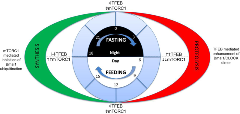
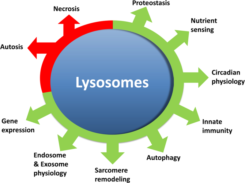
Adaptive responses that counter starvation have evolved over millennia to permit organismal survival, including changes at the level of individual organelles, cells, tissues, and organ systems. In the past century, a shift has occurred away from disease caused by insufficient nutrient supply toward overnutrition, leading to obesity and diabetes, atherosclerosis, and cardiometabolic disease. The burden of these diseases has spurred interest in fasting strategies that harness physiological responses to starvation, thus limiting tissue injury during metabolic stress. Insights gained from animal and human studies suggest that intermittent fasting and chronic caloric restriction extend lifespan, decrease risk factors for cardiometabolic and inflammatory disease, limit tissue injury during myocardial stress, and activate a cardioprotective metabolic program. Acute fasting activates autophagy, an intricately orchestrated lysosomal degradative process that sequesters cellular constituents for degradation, and is critical for cardiac homeostasis during fasting. Lysosomes are dynamic cellular organelles that function as incinerators to permit autophagy, as well as degradation of extracellular material internalized by endocytosis, macropinocytosis, and phagocytosis. The last decade has witnessed an explosion of knowledge that has shaped our understanding of lysosomes as central regulators of cellular metabolism and the fasting response. Intriguingly, lysosomes also store nutrients for release during starvation; and function as a nutrient sensing organelle to couple activation of mammalian target of rapamycin to nutrient availability. This article reviews the evidence for how the lysosome, in the guise of a janitor, may be the "undercover boss" directing cellular processes for beneficial effects of intermittent fasting and restoring homeostasis during feast and famine. © 2018 American Physiological Society. Compr Physiol 8:1639-1667, 2018.


Figures

Figure 1:
Figure 1:. Cardiac metabolism in health and disease:
During normal physiology, the vast majority of myocardial ATP production is from oxidative phosphorylation. Of this, a majority of this is from β-oxidation of fatty acids, with a smaller contribution from glucose oxidation. By contrast, glycolysis, ketone body oxidation and amino acid metabolism contribute very small amounts to overall ATP generation. During pathological states, cardiac myocytes use alternate preferences for energy generation (reviewed in (215) (213); please see text under the section ‘Cardiac energetics, substrate metabolism and pathophysiology under fasting states’ for details). A. Metabolic changes during nutrient deprivation vary depending the duration (ST= short term and LT= long term). During short term fasting, fatty acid oxidation is unchanged while glucose metabolism is suppressed. In addition, marked increases in ketone body oxidation and amino acid metabolism contribute to myocardial energetics. During prolonged fasting, however, lipid oxidation is increased and serves as the main energetic source. B. During ischemic syndromes (I= ischemia and IR= ischemia-reperfusion), energetic choices are driven by oxygen availability. While ischemia is associated with reduction in fatty acid and glucose oxidation, glycolysis serves as a far more significant source of ATP. When reperfused, glycolysis rates decline and glucose oxidation is still suppressed, while fatty acid oxidation increases. In most ischemic syndromes, TCA (tricarboxylic acid cycle)-independent amino acid metabolism is augmented. C. Cardiac myocyte dysfunction in cardiomyopathy is often accompanied by abnormalities in myocardial metabolism and energetics. Prominent among these is a switch from a preference for glucose oxidation as opposed to lipid oxidation. D. During aging, similar to cardiomyopathy, fatty acid oxidation is decreased with increases in glucose oxidation and glycolysis driving myocardial energetics. E. Fatty acid utilization is increased in the diabetic myocardium (with type 2 diabetes due to insulin resistance) with reduction in glucose utilization. The diabetic heart increasingly utilizes ketone bodies as a nutrient source. ’ indicates upregulation, whereas ‘’ indicates downregulation.
Figure 2:
Figure 2:. Mechanisms for execution of autophagy as a lysosomal degradative pathway:
Nutrient deprivation and organelle damage trigger macro-autophagy in cardiac myocytes (reviewed in (68)). Assembly of the phagophore (the initiation of the double membrane) is activated by class III phosphatidylinositol 3-kinase (PtdIns3K) catalyzed generation of phosphatydil inositol 3-phosphate (PtdIns3P) to recruit PtdIns3P-binding proteins. A protein complex is formed comprised of Vps15 homolog phosphoinositide-3-kinase, regulatory subunit 4 (PIK3R4), the Vps34 homolog phosphatidylinositol 3-kinase, catalytic subunit type 3 (PIK3C3), and the Vps30/Atg6 homolog Beclin 1. This interacts with AMBRA1 (not shown) and ATG14, and also contain the BECN1-interacting protein UV radiation resistance associated (UVRAG, not shown) and SH3-domain GRB2-like endophilin B1 (SH3GLB1/Bif-1, not shown) to promote autophagosome formation. Autophagosome formation has been shown to be initiated at multiple sites including the mitochondria-associated membrane, endoplasmic reticulum, the ER-golgi interface and the plasma membrane. PtdIns3P at the ER triggers the recruitment of the PtdIns3P-binding protein ZFYVE1/DFCP1 (zinc finger, FYVE domain containing 1) protein and WD repeat domain, phosphoinositide interacting 2 protein (WIPI2) to from a PtdIns3P-enriched ER-associated structure termed the omegasome for its Ω-like shape. Elongation and expansion of the phagophore membrane takes place by the Atg12–Atg5-Atg16 and Atg8 conjugation systems, which are ubiquitin like conjugation systems. The Atg12–Atg5-Atg16 complex catalyzes the lipidation of Atg8 by Atg4-mediated cleavage followed by covalent linkage to phosphatidylethanolamine. Mammalian Atg8 homologs, namely, the microtubule-associated protein 1 light chain 3 (MAP1LC3/LC3) and GABA (A) receptor-associated protein (GABARAP) subfamily proteins are involved in membrane expansion and also interact with adaptor proteins that bind ubiquitinated proteins (as cargo) to sequester them within the autophagosomes. Adaptor proteins such as p62, NBR1, NDP52, VCP and optineurin interact with ubiquitinated proteins through a ubiquitin-binding domain (UBA) and with LC3 family of proteins via a LC3-interacting region (LIR). After closure of the double membrane, autophagosome-lysosome fusion occurs in a manner dependent upon lysosomal pH (requires acidified lysosomes) and regulated by Rab7, SNARE and HOPS complex proteins. This is followed by intralysosomal degradation of autophagosome contents with recycling of basic building blocks back to the cytosol, completing the process of autophagy. Lysosomes harbor the LYNUS complex on their cytosolic face, which is comprised of mTOR complexed with the lysosomal proton pump, Ragulator, Rag GTPases, GAP (GTPase activating) and GEF (GTP exchange factor) proteins and Rheb GTPase. The LYNUS complex activates mTOR during the nutrient replete state which phosphorylates the TFEB family of transcription factors (reviewed in (162, 188)). Phosphorylated TFEB on lysosome membrane exists in equilibrium with a pool that is bound to cytosolic 14–3-3 proteins. Activated mTOR inactivates autophagy via phosphorylation of ULK1 and ATG13. Nutrient depletion induces the LYNUS complex to unravel, whereby mTOR becomes inactive relieving the constitutive phosphorylation of TFEB. Simultaneously, release of lysosome calcium via activation of the mucolipin channel activates calcineurin, which dephosphorylates TFEB to unmask its nuclear nuclear localization signal. Nuclear TFEB activates transcription of autophagy and lysosome genes, and induces autophagosome formation and lysosome biogenesis to upregulate flux through the macro-autophagy pathway. These lysosomal degradative processes generate nutrients that provide energy and support various metabolic functions (reviewed in (98)), and destroy invading organisms in immune cells via the process of xenophagy and enable antigen presentation to mount an immune response (reviewed in (136).
Figure 3:
Figure 3:. Regulation of Autophagy-Lysosome Machinery:
Various cell-intrinsic and extrinsic inputs regulate the autophagy-lysosome machinery. Organelle damage and impaired protein quality control with dysfunction of the ubiquitin-proteasome system activates selective autophagy to remove the damaged organelles (mitochondria, ER, golgi and lysososomes) and protein aggregates (reviewed in (69)). Aging induces ROS upregulation and accumulation of DNA damage, which induce senescence in cardiac progenitors through telomere attrition and DNA damage (reviewed in (195). While SIRT1 expression is increased, experimental evidence indicates that further stimulation of SIRT1 function enhances autophagy via activation of FOXO1. Serum levels of GDF11, a TGFβ family member, decreases with age in the serum and increasing GDF11 levels beneficially affects age-related cardiac phenotypes. While GDF11 stimulates autophagy in skeletal muscle; its effects on the heart have not been studied. miR-216a increases in the aging heart and is known to downregulate Beclin-1, a protein essential for autophagy induction. Nicotinamide phosphoribosyl transferase (Nampt), a key enzyme in the salvage pathway of NAD+ synthesis in cardiomyocytes is down regulated with aging, and restoring Nampt and NAD+ levels restores autophagic flux in the heart. Multiple transcriptional pathways intricately regulate the autophagy-lysosome machinery (reviewed in (61)). The FOXO family of transcription factors are autophagy activators and are activated by phosphorylation effected by AMPK, JNK and MST1 kinases. However, Akt-mediated phosphorylation of FOXO1 at a different site inactivates FOXO1 and holds it in the cytosol. Activation of the stress sensor ERN1/IRE1α signaling also holds FOXO activity in check. TP53 (tumor protein p53), a transcription factor that regulates cell cycle activates autophagy when localized to the nucleus by directly activating transcriprion of various ATG genes, but inhibits autophagy when in the cytosol. Lysosomal lipases are held in check by mxl3 in C. elegans and by its orthologue MAX in mice. Both are transcriptional repressors that bind to the CLEAR response element where TFEB binds to inhibit lipophagy and lipolysis for generation of energy (reviewed in (98). CREB, or cAMP response element-binding protein is held in check by its interaction with FXR, farnesoid X receptor in the fed state. Fasting induced FXR degradation and binding of CREB to CRTC2, its co-activator to induce TFEB transcription. Fasting also causes LYNUS machinery to deactivate mTOR which results in dephosphorylation and activation of TFEB. TFEB stimulates transcription of autophagy and lysosome pathway genes. Circadian rhythm plays a central role in regulation of the fasting feeding responses and controls autophagy-lysosome machinery. Emerging evidence points to counter-regulation of TFEB family of transcription factors by circadian rhythm regulators either indirectly via mTOR activity or directly via TFEB binding to Per genes (reviewed in (155). Numerous metabolic influences also regulate autophagy in the heart, as shown (and reviewed in (98)). Please see section titled ‘Cardiac energetics, substrate metabolism and pathophysiology under fasting states’ for a detailed discussion of cardiac metabolism under stress. ’ indicates , whereas ‘’ indicates inhibition.
Figure 4:
Figure 4:. Proposed mechanism for the effects of intermittent fasting on LYNUS-TFEB-mTORC1 axis in cardiac myocyte pathophysiology:
Cardiac myocytes in pathological states are likely to be affected by the functioning of the Lysosomal NUtrient Sensing complex (see reference (162) for a detailed review of signaling via the LYNUS complex, and text under the sub-section titled ‘Lysosomes and Nutrient Sensing’). Unopposed activation of mTORC1 as a result of nutrient oversupply, aging, lysosomal dysfunction and diabetes mellitus, is expected to result in a sustained synthetic state. This would result in overload of the ER-translational machinery precipitating ER stress and the unfolded protein response due to accumulation of misfolded entities. This pathological synthetic state is expected to provoke decreased recycling of organelles as well sarcomere renewal and repair. Furthermore, the overall metabolic reserve of the cell would be reduced due to a decrease in the ability of cells to activate catabolic pathways. Similarly, during starvation or with continuous mTOR antagonism, the translation machinery is expected to be suppressed while proteolysis is increased. This would drive breakdown of organelles and sarcomeres with suppression of organelle and sarcomere biogenesis. In addition, progressive depletion of cellular components by catabolic pathways is expected to result in a similar decrease in metabolic reserve. In contrast, intermittent fasting drives cyclic activation of both TFEB and mTORC1, which is expected to optimize protein turnover. This is accompanied by decreased ER stress, increased organelle turnover, where accelerated organelle/sarcomere breakdown is accompanied by increase biogenesis and optimal availability of cellular components and metabolic pathways to increase metabolic reserve. As compared to cardiac myocyte dysfunction seen with sustained activation of either mTORC1 or TFEB, acceleration of the cyclic activation of mTORC1 and TFEB is expected to results in cardiac myocyte rejuvenation. Similar mechanisms are proposed to drive the beneficial effects of exercise, intermittent rapamycin, circadian feeding and sirtuin action.
Figure 5:
Figure 5:. Proposed LYNUS-TFEB-mTORC1 cues to entrain the circadian rhythm.
Dietary factors have been noted to affect circadian physiology. In this proposed mechanism, diurnal variation in feeding behavior is accompanied by the effect of mTORC1 and TFEB on the Bmal1 and Clock heterodimer resulting in synchronization of the circadian clock with nutrient supply. High TFEB levels at waking (“Day” in diurnal species) enhance the Bmal1/CLOCK heterodimer, thus driving circadian genes. In contrast, peak mTORC1 activation at the onset of “night” phase, when feeding is complete affects Bmal1 ubiquitination and alters the circadian gene expression, coincident with the peak synthetic phase. As opposed to these two phases, the transition from synthesis to proteolysis (and vice versa) at ‘0’ and ‘12’, is also accompanied by combined effects of TFEB and mTORC1 on the Bmal1/CLOCK proteins. By adhering to a fixed pattern, it may be thus possible to synchronize the metabolic and biologic clocks, and thus maximize cellular repair and renewal while optimizing metabolism. Please also see text under the section titled ‘Interplay of lysosome function with the metabolic circadian axis’.
Figure 6:
Figure 6:. Lysosomal regulation of life and death
Lysosomes play multiple salutary roles as well as participate in orchestrating cell death programs. Necrosis and autosis (or autophagic cell death) are both characterized by lysosomal digestion of cellular contents. Please see reference (67) for a comprehensive review of the role of lysosomes and autophagy in cell death. In contrast to this destructive role, lysosomes play critical roles in both synthesis and non-proteasomal breakdown of proteins. By virtue of the Lysosomal NUtrient Sensing mechanisms, they control cellular metabolic choices as well as circadian physiology. Lysosomes are also vital for cellular innate immunity against pathogens as well as dysfunctional organelles, sarcomeres and even, pathologically aggregated proteins. Through effects on TFEB and mTORC1, lysosomes can control gene expression as well as the translational machinery. Finally, processing of both intracellular and extracellular messages, via exosomes and endosomes, is regulated via lysosomal function.

References

    1. Abu-Remaileh M, Wyant GA, Kim C, Laqtom NN, Abbasi M, Chan SH, Freinkman E, and Sabatini DM. Lysosomal metabolomics reveals V-ATPase- and mTOR-dependent regulation of amino acid efflux from lysosomes. Science 358: 807–813, 2017. - PMC - PubMed
    1. Acosta-Rodriguez VA, de Groot MHM, Rijo-Ferreira F, Green CB, and Takahashi JS. Mice under Caloric Restriction Self-Impose a Temporal Restriction of Food Intake as Revealed by an Automated Feeder System. Cell Metab 26: 267–277 e262, 2017. - PMC - PubMed
    1. Adeva-Andany MM, Gonzalez-Lucan M, Donapetry-Garcia C, Fernandez-Fernandez C, and Ameneiros-Rodriguez E. Glycogen metabolism in humans. BBA Clin 5: 85–100, 2016. - PMC - PubMed
    1. Ahmet I, Wan R, Mattson MP, Lakatta EG, and Talan M. Cardioprotection by intermittent fasting in rats. Circulation 112: 3115–3121, 2005. - PubMed
    1. Ahmet I, Wan R, Mattson MP, Lakatta EG, and Talan M. Cardioprotection by intermittent fasting in rats. Circulation 112: 3115–3121, 2005. - PubMed

Publication types

LinkOut - more resources